(UroToday.com) The Advanced Prostate Cancer Consensus Conference 2021 meeting session discussing PSMA in diagnostics and therapy included a presentation by Dr. Derya Tilki discussing background information of PSMA imaging and providing context for the subsequent presentations in this session. Dr. Tilki started by highlighting that PSMA is a transmembrane protein with enzymatic function as a folate hydrolase-carboxypeptidase, with a short cytoplasmic domain, a hydrophobic transmembrane region, and a large extracellular domain. PSMA is highly expressed on prostate epithelial cells and is strongly upregulated in prostate cancer (~1000-fold higher expression compared with other tissues). Interestingly, PSMA was recognized as a target >3 decades ago. The short intracellular domain contains a binding site that can be targeted with 7E11 antibodies, and in 1996 ProstaScint (111In-labelled 7E11) was approved by the FDA (the first commercial monoclonal PSMA antibody), but only reaches the cells that are already in apoptosis:

Subsequently, antibodies and small molecules that target the extracellular domain of PSMA were developed.
Physiologically, there is high-intensity PSMA activity seen in the lacrimal, parotid, and submandibular glands, and it may also be seen in the small bowel (primarily the duodenum). Of note, the highest intensity uptake is seen in the kidneys, ureters, and bladder, given that the compound is renally excreted. There is also moderate-intensity uptake seen in the liver and spleen, with low uptake in the parasympathetic ganglia. Dr. Tilki notes that most of the non-prostate cancer-related uptake of PSMA PET has low intensity or is non-focal, in contrast to the focal and intense uptake in prostate cancer lesions.
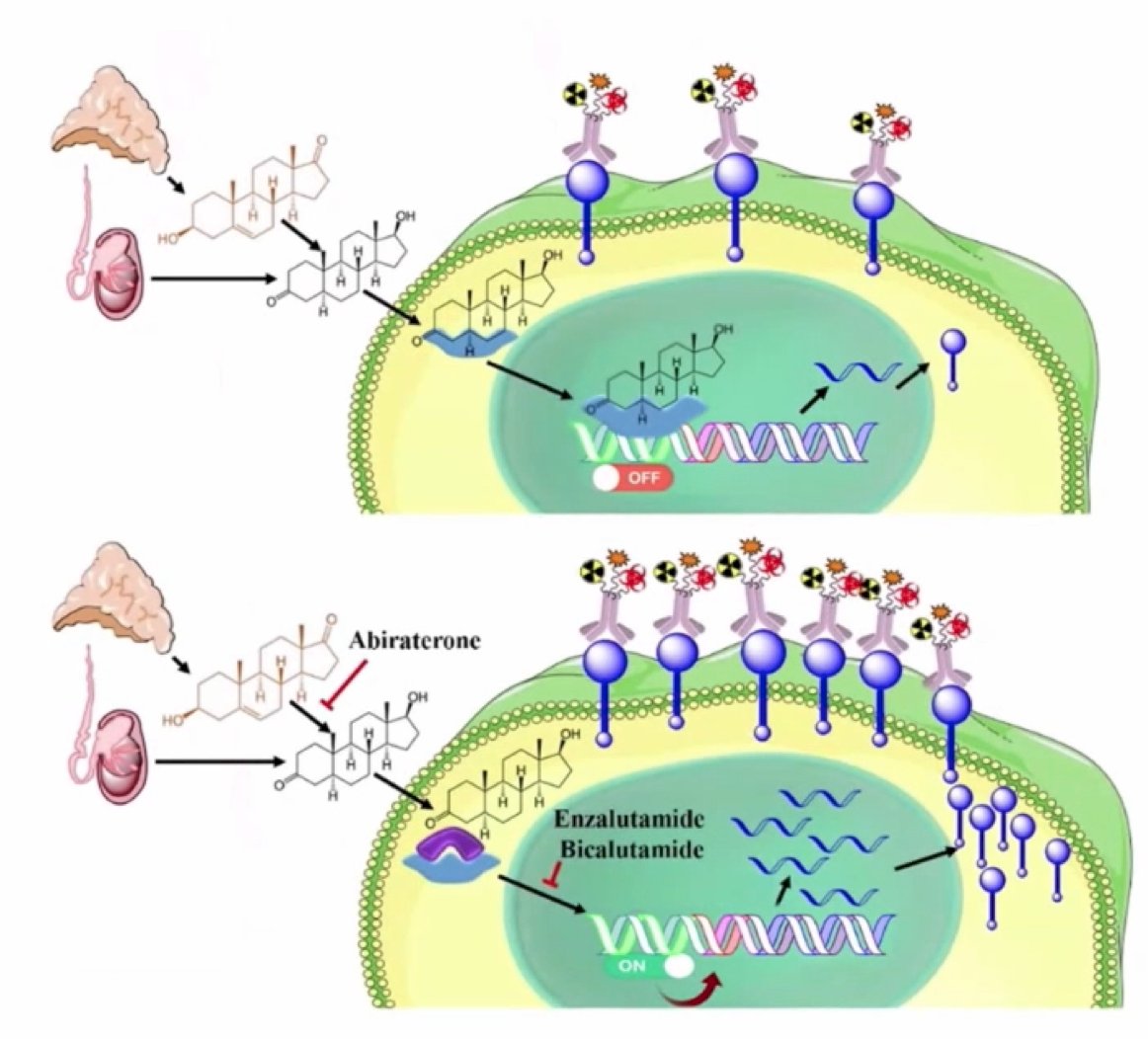
Treatment with ADT results in upregulation in PSMA expression. The folate hydrolase 1 gene encodes the PSMA protein, and folate hydrolase 1 gene expression is suppressed by androgens. In preclinical models, ADT increases expression of PSMA in both castration-sensitive and castration-resistant prostate cancer:1
In an early case report of a patient with castration-sensitive prostate cancer (Gleason score 10) before and four weeks after treatment with ADT, there was a 7-fold increase in PSMA uptake after the initiation of ADT. Additionally, 13 of 22 lesions in this patient were visualized on PSMA PET only after treatment with ADT. In a study by Emmett et al.,2 serial 68Ga-PSMA PET scans were obtained at baseline and on days 9, 18, and 28 among 8 men with mHSPC and 7 men with mCRPC. Among the men with mHSPC, a median 30% reduction in SUVmax was recorded by day 9, after which PSMA response heterogeneity was noted, with persistently high or increasing SUVmax in 37.5% and reduction in 62.5%. Among men with mCRPC, all patients demonstrated an increase in SUVmax, with the increase seen at day 9 plateauing by day 28. Since PSMA expression on PSMA-PET is related to both the number of cells expressing PSMA and target level expression, initial reduction in the castration-sensitive setting may be in part attributable to tumor shrinkage in response to ADT as opposed to reduced PSMA expression per cell.
Dr. Tilki also emphasized that there is intra- and interpatient heterogeneity of PSMA expression. In a study by Paschalis et al,3 they noted marked intratumor heterogeneity of membranous PSMA expression, with foci containing no detectable PSMA, observed in all membranous PSMA expressing castration-sensitive prostate cancer (100%) and 37 (84%) metastatic CRPC biopsies. Heterogeneous intra-patient membranous PSMA expression between metastases was also observed, with the lowest expression in liver metastases. Higher levels of membranous PSMA expression were associated with higher Gleason grade and worse overall survival.
Dr. Tilki concluded her presentation with the following summary points:
- PSMA is predominantly localized to the prostate, kidneys, salivary glands, nervous system glia, and the small bowel
- PSMA expression appears to be progressively increased in higher-grade tumors, and in castration-resistant and metastatic prostate cancer
- Preclinical studies have shown that ADT increases expression of PSMA in both castration-sensitive and castration-resistant prostate cancer
- PSMA expression demonstrates marked inter- and intra-patient heterogeneity
Presented by: Derya Tilki, MD, Department of Urology, Martini-Klinik Prostate Cancer Center, University Hospital Hamburg-Eppendorf, Hamburg, Germany
Written by: Zachary Klaassen, MD, MSc – Urologic Oncologist, Assistant Professor of Urology, Georgia Cancer Center, Augusta University/Medical College of Georgia, @zklaassen_md on Twitter during the 2021 Advanced Prostate Cancer Consensus Conference, Saturday, October 9, 2021.
References:
- Bakht MK, Oh SW, Youn H, et al. Influence of androgen deprivation therapy on the uptake of PSMA-targeted agents: Emerging opportunities and challenges. Nucl Med Mol Imaging. 2017;51(3):202-211.
- Emmett L, Yin C, Crumbaker M, et al. Rapid modulation of PSMA expression by androgen deprivation: Serial 68Ga-PSMA-11 PET in men with hormone-sensitive and castrate-resistant prostate cancer commencing androgen blockade. J Nucl Med. 2019;60(7):950-954.
- Paschalis A, Sheehan B, Riisnaes R, et al. Prostate-specific membrane antigen heterogeneity and DNA repair defects in prostate cancer. Eur Urol. 2019;76(4):469-478.