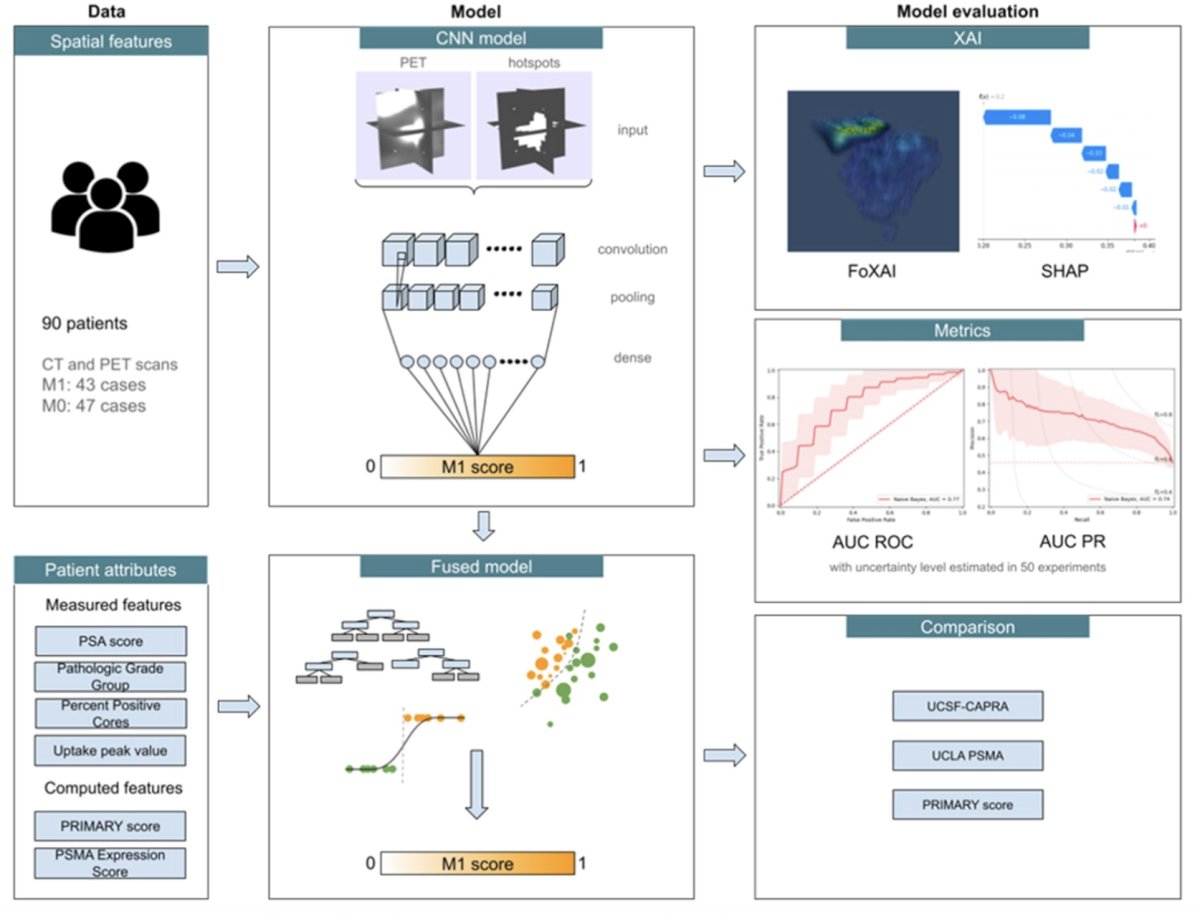
(UroToday.com) The 2024 GU ASCO annual meeting featured a prostate cancer session and a presentation by Dr. Jesus Eduardo Juarez discussing a convolutional neural network model using intraprostatic patterns of 18F-DCFPyL uptake in PSMA PET images to predict synchronous metastases. 18F-DCFPyL is a PSMA-targeted imaging agent that provides whole-body staging of prostate cancer. As such, there may be untapped data contained within 18F-DCFPyL PET images that can be used for prognostic evaluation. Dr. Juarez and colleagues developed convolutional neural network models using inputs from the entire prostate on 18F-DCFPyL images along with auto-segmented hot spots within the of primary prostate cancer tumor to predict synchronous metastases and compared the models against established models built from clinicopathologic data.
This study included 91 Veterans with de novo prostate cancer who were imaged with 18F-DCFPyL PSMA PET/CT for initial staging (46% with metastatic disease). The 18F-DCFPyL images of the prostate were analyzed using aPROMISE (PYLARIFY AI), which autosegments, localizes, and quantifies disease on PSMA PET. The segmentation of the prostate were used to map the 18F-DCFPyL PET image of the prostate. Both the entire prostate, as well as aPROMISE defined hot spots were used as inputs for the convolutional neural network, where, according to attention map analysis, the hotspot information helps the network understand the location and extent of tumors:

The dataset was then split into training, validation, and test sets using stratified random sampling. The area under ROC (AUC) curve was computed to determine the performance of the model in predicting the presence of metastases and the test predictions were compared with ground truth (M1). The training was repeated 50 times and the best-performing experiment was identified. Prediction scores from UCSF-CAPRA and UCLA PSMA risk calculator were used as comparators.
The attention map for the convolutional neural network trained on 18F-DCFPyL PSMA PET/CT imaging alone demonstrates that the convolutional neural network model failed to focus on lesion-rich areas:

However, adding the aPROMISE-defined lesions as a separate channel input allows the convolutional neural network model to focus on regions of tumor within the 18F-DCFPyL PSMA PET/CT image:

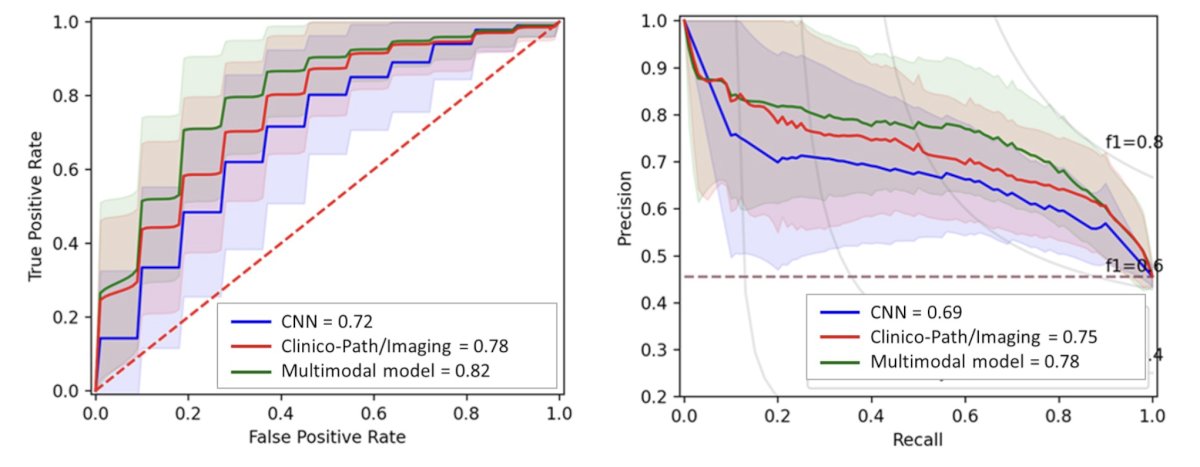
The convolutional neural network model was comparable to the best performing model based on clinicopathologic and measurable imaging data. Models were complimentary and a combined model performed best (AUC 0.82):

AUC and Precision-Recall curves for the convolutional neural network model alone, clinicopathologic and measurable imaging data, and combined model are shown below:

The AUC using UCSF-CAPRA score and UCLA PSMA risk calculator (detecting any upstaging on PET) in this dataset were 0.73 and 0.75, respectively.
Dr. Juarez concluded his presentation discussing a convolutional neural network model using intraprostatic patterns of 18F-DCFPyL uptake in PSMA PET images to predict synchronous metastases with the following take-home points:
- The convolutional neural network based model using 18F-DCFPyL imaging demonstrates that synchronous metastases can be predicted from intraprostatic 18F-DCFPyL uptake patterns alone with a predictive accuracy in this dataset that is at least comparable to published prediction models based on clinicopathologic features
- This study raises the hypothesis that 18F-DCFPyL convolutional neural network-based models could be developed to prognosticate metastatic progression
Presented by: Jesus Eduardo Juarez, MD, Department of Radiation Oncology, UCLA, Los Angeles, CA
Written by: Zachary Klaassen, MD, MSc – Urologic Oncologist, Associate Professor of Urology, Georgia Cancer Center, Wellstar MCG Health, @zklaassen_md on Twitter during the 2024 American Society of Clinical Oncology Genitourinary (ASCO GU) Cancers Symposium, San Francisco, CA, January 25th – January 27th, 2024


