(UroToday.com) The 2024 GU ASCO annual meeting featured a prostate cancer session and a presentation by Dr. Lena Unterrainer discussing low- and high-volume disease in mHSPC, from CHAARTED to PSMA-PET. High-volume disease and low-volume disease definitions are based on conventional imaging (CT + bone scan) according to CHAARTED.
Additionally, volume of disease is associated with overall survival and used for treatment decisions in metastatic hormone sensitive prostate cancer (mHSPC) patients. However, it remains unknown how these definitions transfer to PSMA-PET. The objective of this study was to transfer CHAARTED definitions of high volume disease and low volume disease (as assessed by conventional imaging) to PSMA PET/CT.
mHSPC patients from 5 international sites who underwent PSMA PET + bone scan within a maximum time interval of 100 days and without any new treatment in between were retrospectively included. CHAARTED stratification into high-volume disease/low-volume disease was applied to bone scan, CT, MRI, and PSMA PET. High-volume disease was defined by the presence of visceral metastases and/or ≥ 4 bone metastases (with ≥ 1 beyond spine/pelvis). EXINIbone 3.4 (EXINI Diagnostics) was used for the automated bone scan index and the number/localization of lesions on bone scan. The whole body PSMA PET positive tumor volume as obtained using a semi-automatic thresholding method on Affinity 3.0.2.
There were 67 pairs of PSMA PET/CT + bone scans included for patients that had not had ADT with a median PSA of 208.4 ng/mL (range 0.2-2,840):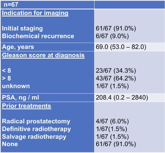
Based on conventional imaging, 25.4% of patients had conventional imaging high-volume disease and 74.6% of patients had conventional imaging low-volume disease. Based on PSMA PET, 40.3% of patients had PSMA high-volume disease and 35.8% had PSMA low-volume disease, and 23.9% of patients had no PSMA PET positive lesion or only local / N1-disease. Upshift and downshift from conventional imaging to PSMA-PET occurred in 27 of 67 (40.3%) patients, including 11 of 67 (16.4%) patients that were upstaged and 16 of 67 (23.9%) patients that were downstaged by PSMA PET: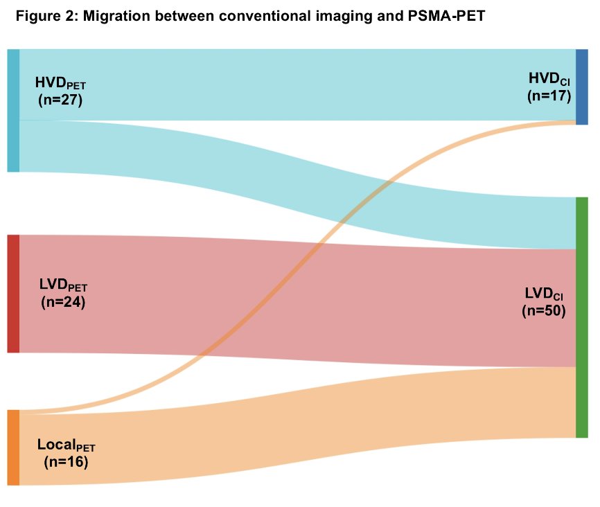
The downshifted patients had no PSMA PET-positive lesion or only in the prostate fossa while conventional imaging was M1 disease. The mean whole-body PSMA PET tumor volume and automated bone scan index was 243 ml (range: 0–3734.0) and 1.1% (range: 0–11.7), respectively. A PSMA tumor volume of 107 mL was the optimal cut off between CHAARTED low volume and high volume disease (misalignment of 20.9%).
Dr. Unterrainer concluded this presentation discussing low and high volume disease in mHSPC, from CHAARTED to PSMA-PET with the following take-home points:
- PSMA-PET based volume of disease definitions may be redefined by correlation with patients’ outcome
- Will higher accuracy on PSMA-PET (compared to conventional imaging) lead to improved outcomes?
- The impact of the localization/volume of bone metastases versus PSMA tumor volume for new volume of disease definitions remains to be defined
Presented by: Lena Unterrainer, MD, Ahmanson Translational Theranostics Division, Department of Molecular and Medical Pharmacology, UCLA, Los Angeles, CA
Written by: Zachary Klaassen, MD, MSc – Urologic Oncologist, Associate Professor of Urology, Georgia Cancer Center, Wellstar MCG Health, @zklaassen_md on Twitter during the Genitourinary (GU) American Society of Clinical Oncology (ASCO) Annual Meeting, San Francisco, CA, Thurs, Jan 25 – Sat, Jan 27, 2024.