Contrast-enhanced ultrasound (CEUS) uses microbubble contrast agents and is sensitive to detect low blood flow and tissue perfusion information (figure 1). Its advantage over CT/MRI includes no ionizing radiation, no nephrotoxicity, and low cost. The Superb Microvascular Imaging (SMI) has been FDA-cleared since 2014. It is a novel Doppler technique which detects lower blood flow velocities, higher resolution, and sensitivity, and does not require a contrast agent. It also has fewer motion artifacts, with higher frame rates (>50 Hz) and has been investigated in multiple applications.

The Aplio i800 scanner was used in this study, with several modalities used for the study including:
- CDI – Color Doppler imaging
- PDI – Power Doppler imaging
- cSMI – Color superb microvascular imaging
- mSMI – Monochrome superb microvascular imaging
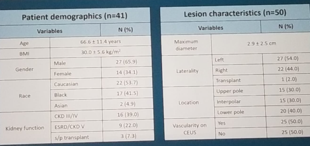
Patient demographics and lesion characteristics are shown in table 1. The sensitivity, specificity, accuracy, positive and negative predictive values of the various ultrasound modalities are shown in table 2. The receiver operating characteristics (ROC) curve for vascularity of IRM for the different modalities is shown in figure 2, with an AUC of 0.63, 0.62, 0.68, and 0.68 for CDI, PDI, cSMI, and mSMI, respectively, with cSMI demonstrating the best results.
Table 1 – Basic demographic data and lesion characteristic

Table 2- The sensitivity, specificity, accuracy, positive and negative predictive values of the various ultrasound modalities:

Figure 2 – ROC – AUC curve for the vascularity of indeterminate renal masses for the different ultrasound modalities:

The limitations of this study include lack of final pathology or contrast-enhanced cross-sectional imaging, the fact that multiple radiologists read clinical CEUS images, limited reader experience evaluating this new technology, and a wide variety of clinically indeterminate renal masses.
Dr. Leong concluded his talk, stating that this very early preliminary data suggest that SMI is potentially superior to CDI and PDI in detecting micro-vascularization in IRM. SMI may be a diagnostic tool for stratification of IRM in 40% of cases, but at this point, CEUS is still needed for confirmation of vascularity.
Presented by: Joon Yau Leong, MD, Department of Urology, Thomas Jefferson University
Written by: Hanan Goldberg, MD, Urologic Oncology Fellow (SUO), University of Toronto, Princess Margaret Cancer Centre @GoldbergHanan at the American Urological Association's 2019 Annual Meeting (AUA 2019), May 3 – 6, 2019 in Chicago, Illinois


