(UroToday.com) At the annual American Urological Association conference in Chicago, moderated poster session 26 was about recent advancements in surgical technology, specifically regarding the simulation of instrumentation. Dr. Amy Reed and her team from Vanderbilt University Medical Center explored recent advancements in computer vision, a field of computer science that focuses on enabling computers to identify and understand objects in images and videos. The team sought to apply this technology to upper urinary tract tumors during ureteroscopies. Using this technology, they hope to improve tumor visualization and automatically detect urothelial tumors during real time ureteroscopy.
Dr. Reed et. al used 8 separate surgical videos of urothelial tumor diagnosis and ablation via digital ureteroscopies. Two key aspects needed to be identified from each video, initial tumor identification and tumor ablation. Frames were annotated by one urologist to identify the key aspects of all 1097 frames extracted from the videos. 80% of these frames were used to train the computer vision model, the remaining 20% were reserved as a test set to automatically segment the model. Once the machine was automatically segmented it was compared to the manual annotation for accuracy (Figure 1). Dr. Reed and her team were able to correctly identify 23 different tumors that were ablated using a holmium laser. The model was able to correctly identify with an accuracy of 0.95 (Figure 2).
The initial use of this technology proves to be successful in identifying tumors, however, this is based on videos that have been previously recorded. Dr. Reed concluded her presentation by saying that further developing this technology can ultimately result in applying this technology in real time which may be used to improve ablation rates and be used for better tracking and surveillance. Following her presentation, one audience member asked what is being used as an irrigation fluid water or saline as one may provide a better visualization. To which she answered with ease that saline was being used the irrigate. Another question arose as to what is being used as the “ground truth” for the program. Dr. Reed replied that since these were done in a retrograde fashion, visualization from the urologist was the determinant of what were the correct structures to identify. Dr. Reed and her team have developed promising technology and if further developed may be able to be used in real time during ureteroscopies, an advancement that may help urologists worldwide better understand the characteristics of tumors while operating.
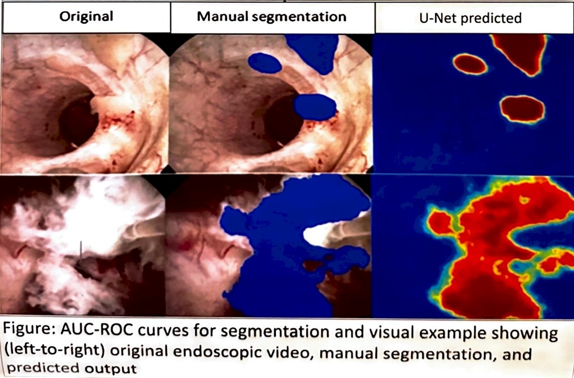
Figure 1. Shows how Dr. Reed and her team took videos from ureteroscopies, manually segmented them and how the computer vision identified them.
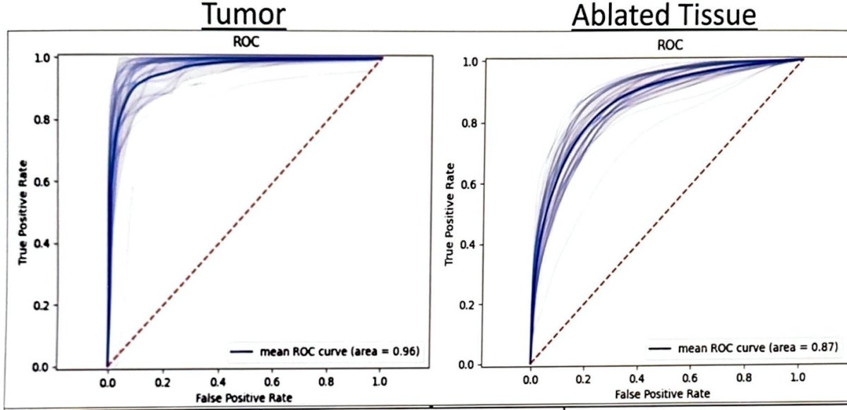
Figure 2. AUC-ROC curves for segmentation demonstrates the computer visions promising accuracy.
Presented by: Amy Reed M.D., Vanderbilt University Medical Center
Written by: Paul Piedras, B.S., University of California, Irvine, @piedras_paul on twitter, during the 2023 American Urological Association (AUA) Annual Meeting, Chicago, IL, April 27 – May 1, 2023