Dr. Volpe emphasized that, while robotic-assisted partial nephrectomy can be technically challenging, the goal should always be to recapitulate open surgical principles including preparation of the renal hilum, mobilization of the kidney, clamping of the hilum, tumor excision, and renorrhaphy. He emphasized that, even when non-ischemic resection is planned, hilar mobilization is critical for the safe performance of the case.
Dr. Volpe described and provided videos of, both a standard approach to the hilum, working from caudal to cranial, as well as a top-down approach (on the right) in which the peritoneum overlying the kidney below the liver is incised, allowing access to the vena cava which may then be followed down to the hilum.
Recapitulating open approaches, Dr. Volpe emphasized that the renal artery can be identified for right-sided resections in the interaortocaval space, particularly when full arterial clamping is required, particularly where there is early branching of the artery. Such an approach requires extensive mobilization of the colon and duodenum.
Dr. Volpe then emphasized the significant variability in renal hilar vasculature. In approaching these cases, he highlighted the value of 3-dimensional reconstructions and 3-D printed models. This is of particular value, in his view when selective clamping is planned. Taking this a step further, Dr. Volpe discussed augmented reality in which there are overlay and synchronization of virtual radiographic images with live endoscopic views in the operative field. This has particular value when attempting to dissection secondary and tertiary branches of the vessels.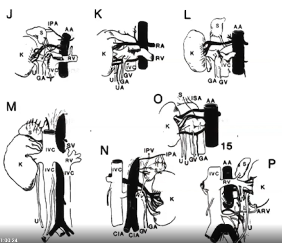
Dr. Volpe then emphasized that the preparation of the hilum is a potentially dangerous step that can cause a sudden, potentially life-threatening situation. As a result, it is necessary to be prepared for urgent conversion to an open approach.
To guide hilar dissection, Dr. Volpe highlighted the value of ICG fluorescence. The use of intravenous indocyanine green IV with the da Vinci Fluorescence Imaging Vision System (Firefly Fluorescence Imaging) assists with the identification of vascular anatomy, as highlighted in the figure below.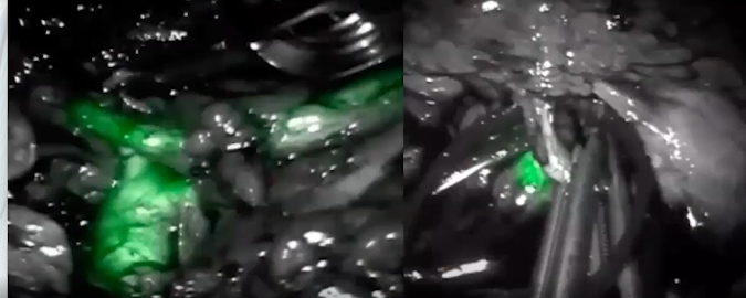
In addition to this, fluorescence can assess with identification of the efficacy of selective arterial clamping and test parenchymal perfusion following renorrhaphy.
Presented by: Alessandro Volpe, MD, Associate Professor of Urology, Urologic Oncologist, Chairman Division of Urology -University of Eastern Piedmont, Maggiore della Carità Hospital, Novara, Italy
Written by: Christopher J.D. Wallis, MD, Ph.D., FRCSC, Instructor in Urology, Vanderbilt University Medical Center, Nashville, Tennessee @WallisCJD on Twitter at the 18th Meeting of the EAU Section of Oncological Urology (ESOU21), January 29-31, 2021