(UroToday.com) The 2023 Society of Nuclear Medicine and Molecular Imaging (SNMMI) Annual Meeting held in Chicago, IL between June 24th and 27th, 2023 was host to a prostate cancer session. Dr. Kilian Kluge presented the results of an analysis evaluating the effects of anti-hormonal treatment status on ⁶⁸Ga-PSMA-HBED-CC positron emission tomography (PET) biodistribution in prostate cancer patients.
Prostate-specific membrane antigen (PSMA) expression has been shown to be modulated by androgen deprivation therapy (ADT) exposure. Although a clear consensus is lacking, it appears that short-term ADT exposure is associated with increased PSMA expression (‘PSMA flare’),1,2 whereas long-term ADT exposure is associated with decreased PSMA expression.3 Most studies to date have focused on the hormonal influence on PSMA expression in tumoral tissues, with influence on non-tumor tissues only investigated in short-term settings. This is of utmost clinical relevance given that adverse effects and subsequent dose limitations for PSMA radioligand therapy may be caused by PSMA expression, retention, and excretion in these non-tumoral tissues.
The study objective was to retrospectively compare 68Ga-PSMA-HBED-CC PET uptake values of the salivary gland, kidneys, liver, and spleen in patients receiving ADT versus those with no prior history of ADT exposure. To this end, the authors queried their institutional database and included a total of 112 patients (56 receiving ADT and 56 controls). Patients receiving androgen receptor signaling inhibitors (ARSI; e.g., abiraterone, enzalutamide, etc.) were excluded from the analysis. Patients with a metabolic tumor volume (MTV) >25 cm3 were excluded, given that intense tracer uptake in such tumors would ‘deplete’ the available remaining tracer to be taken up by non-tumor tissue. The 56 patients in each of the 1st generation ADT and control groups were matched by glomerular filtration rate and MTV.

Organ delineation was performed using automatic, objective organ segmentation (MOOSE),4 followed by manual adjustments of:
- Submandibular glands
- Spleen
- Liver
- Kidney
The baseline patient demographics are summarized in the table below. The mean age was 70 years. The mean PSA was higher in the control arm (13 versus 7.1 ng/ml). The MTV was conversely higher in the ADT group (5 versus 3.3 cm3). The majority of patients in the ADT arm were exposed to ADT for >6 months (73.2%).

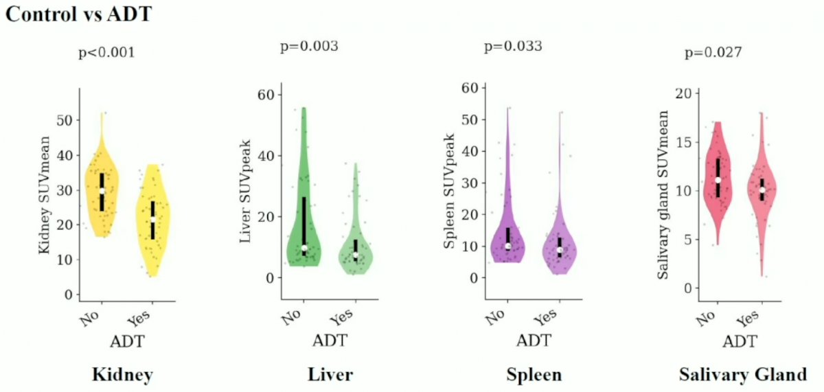
Patients in the ADT arm had significantly lower PSMA expression, quantified via SUVmean or SUVpeak, in the kidneys, liver, spleen, and salivary glands, as summarized in the plots below.
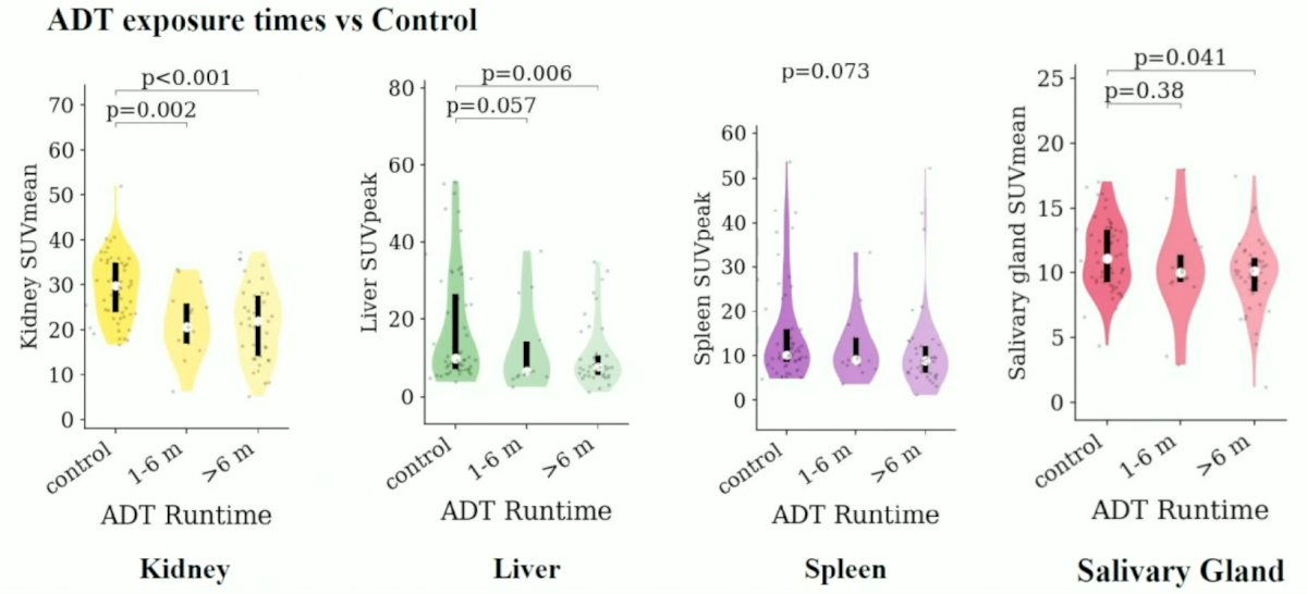
When stratified by ADT exposure duration (1-6 months versus >6 months), the authors noted that PSMA expression was consistently lower in the ADT >6 months group, compared to controls, in the kidneys, liver, spleen, and salivary glands. Compared to controls, PSMA expression in the ADT 1-6 months group was lower in the kidney (p=0.002) and potentially the liver (p=0.057).
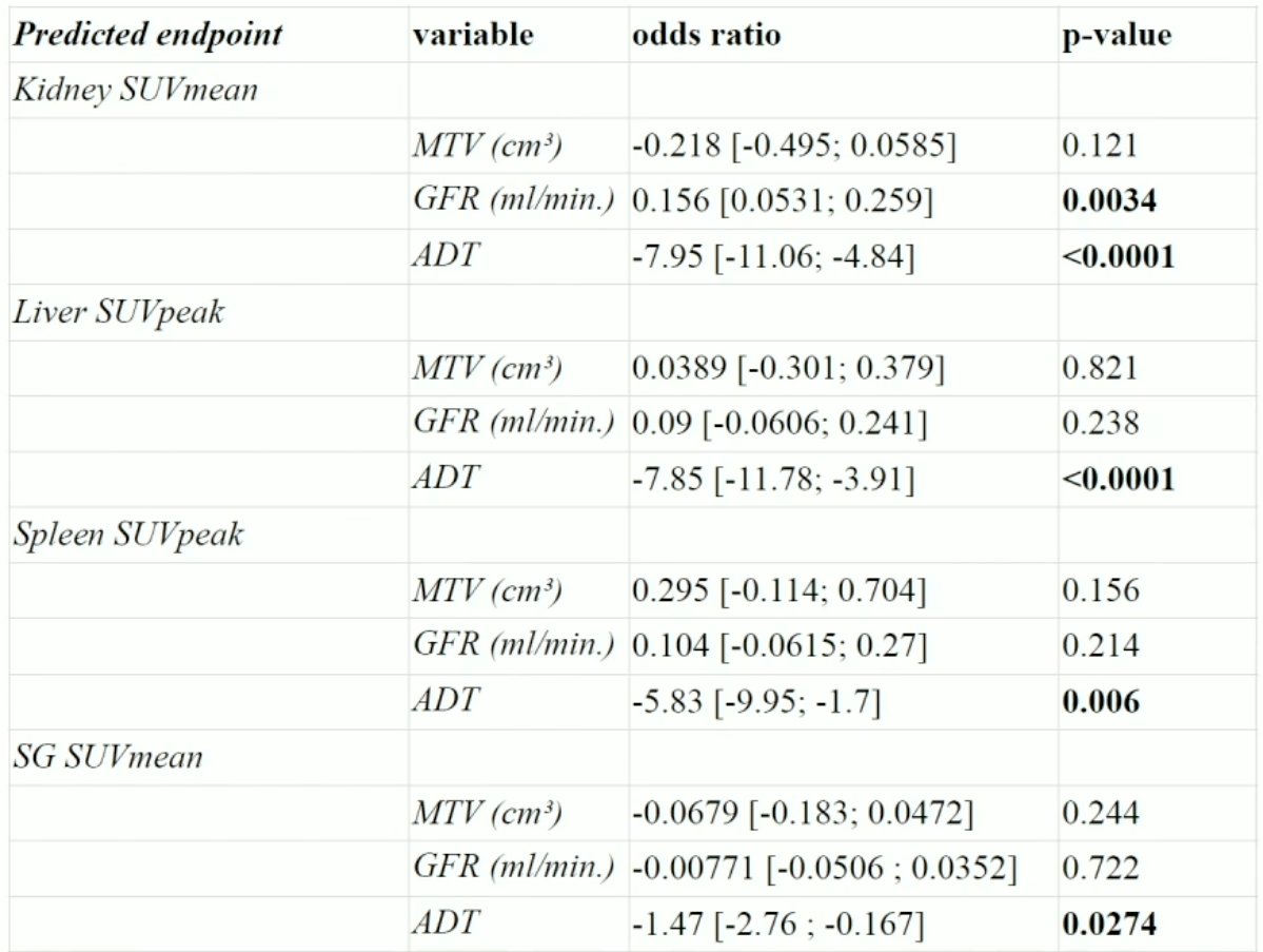
On multivariable analysis adjusted for GFR and MTV, the authors demonstrated that ADT exposure was significantly associated with decreased SUVmean or SUVpeak in the kidneys, liver, spleen, and salivary glands.
Dr. Kluge concluded that sustained ADT exposure is associated with decreased renal, hepatic, splenic, and salivary glandular uptake values. This suggests systemic hormonal modulation of PSMA expression and may have implications for side effect minimization strategies. The major limitations of this study were the single center, retrospective nature of the analysis. No dosimetric measures were used in this study. Future studies will need to evaluate the effect of ARSI use on PSMA expression in non-tumoral tissue and whether prolonged ADT/ARSI exposure prior to radioligand therapy is associated with decreased side effects.
Presented by: Kilian Kluge, MD, Resident Physician, Division of Nuclear Medicine, Medical University of Vienna, Vienna, Austria
Written by: Rashid K. Sayyid, MD, MSc – Society of Urologic Oncology (SUO) Clinical Fellow at The University of Toronto, @rksayyid on Twitter during the 2023 Society of Nuclear Medicine and Molecular Imaging (SNMMI) Annual Meeting, Chicago, IL, Sat, June 24 – Tues, June 27, 2023.
References:- Emmett L, et al. Rapid Modulation of PSMA Expression by Androgen Deprivation: Serial 68Ga-PSMA-11 PET in Men with Hormone-Sensitive and Castrate-Resistant Prostate Cancer Commencing Androgen Blockade. J Nucl Med , 2019;60(7):950-4.
- Hope T, et al. 68Ga-PSMA-11 PET Imaging of Response to Androgen Receptor Inhibition: First Human Experience. J Nucl Med, 2017;58(1):81-4.
- Ashfar-Oromieh A, et al. Performance of [68Ga]Ga-PSMA-11 PET/CT in patients with recurrent prostate cancer after prostatectomy—a multi-centre evaluation of 2533 patients Eur J Nucl Med Mol Imaging, 2018;48:2925-34.
- Sundar LKS, et al. Fully-automated, semantic segmentation of whole-body 18F-FDG PET/CT images based on data-centric artificial intelligence. J Nucl Med, 2022;63(12):1941-8.