(UroToday.com) The 2023 Society of Nuclear Medicine and Molecular Imaging (SNMMI) Annual Meeting held in Chicago, IL between June 24th and 27th, 2023 was host to a prostate cancer session. Dr. Mahbod Jafarvand presented the results of a single center, retrospective analysis evaluating PSMA-PET/CT for prostate cancer patients following focal therapy.
While PSMA-PET/CT has been approved for the staging of patients in the pre-treatment and biochemically recurrent settings, its potential value/performance in patients who had previously underwent focal therapy remains unclear. To date, there are only a limited number of published articles evaluating PSMA-PET/CT following focal therapy, which includes high intensity focused ultrasound (HIFU), irreversible electroporation (IRE), photodynamic therapy, cryoablation, and laser therapy.
To this end, the authors conducted a retrospective, single center analysis, which included patients who received focal therapy for prostate cancer, subsequent 68Ga-PSMA-11 PET with contrast-enhanced CT obtained at 60+ minutes (standard) and 90+ minutes (delayed) images, and no intervening treatment in the interval between focal therapy and PET images acquisition.
The primary study objectives of this study were to evaluate the detection/positivity rate per patient and location of the findings, evaluated via miTNM staging, as well as the diagnostic performance per lesion. Secondary objectives included evaluating inter-reader agreement for prostate positivity rate and positive predictive value per patient.
All images were interpreted by three independent, highly-experienced blinded readers. Since there are no strict guidelines for positivity rate following focal therapy, interpretation was performed based on readers’ experience. A per-region (T, N, M) majority rule was applied for positivity rate. The inter-reader agreement for the prostate positivity rates were calculated using Fleiss’ Kappa value.
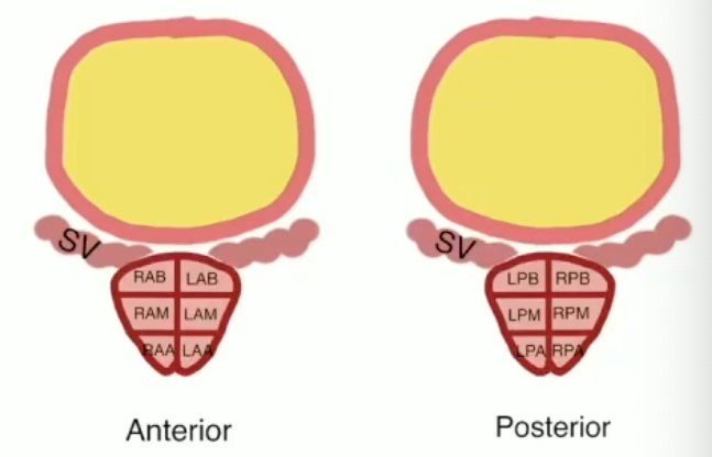
In a subcohort of patients with a biopsy and MRI available within three months of the PSMA-PET/CT acquisition, the authors calculated the diagnostic accuracy parameters on a per-patient and per-segment level. Twelve prostatic segments were defined and matched per pathology report (anterior/posterior and right/left: apex, mid-gland, base). The SUVmax of suspected lesions were measured on standard and delayed acquisition images. The MRI images were analyzed by a single blinded radiologist. Clinically significant prostate cancer (csPCa) was defined as Grade Group two or worse disease.
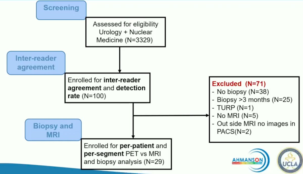
The investigators screened 3,329 charts and identified 100 patients who underwent focal therapy followed by PSMA-PET/CT. These 100 patients were included in the analysis for inter-reader agreement and detection rate. Of these 100 patients, 29 had a repeat biopsy and an MRI within three months of the PET/CT and were enrolled in the per-patient and per-segment diagnostic accuracy analysis cohort.
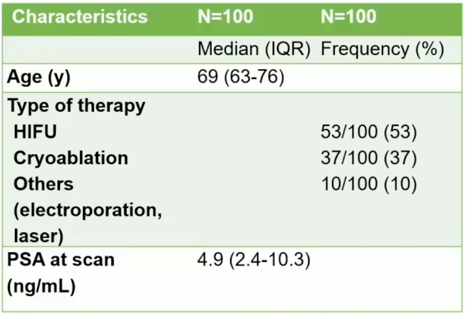
The median patients age of the 100-patient cohort was 69 years (IQR: 63 - 76). The choice of focal therapy was HIFU for 53%, cryoablation for 37%, and another modality in the remaining 10%. The median PSA at the time of the PSMA-PET/CT scan was 4.9 ng/ml (IQR: 2.4 -10.3 ng/ml).
Among the 100 patients, a positive PSMA-PET/CT was noted in 85% of patients, with all having positive prostatic lesions. Extraprostatic extension and seminal vesicle invasion were present in 3% and 26% of patients, respectively. Positive pelvic lymph nodes were noted in 17% and distant metastases in 9%. The SUVmax on standard (60 minute) and delayed (90 minute) acquisition imaging was 7.6 and 9.0, respectively.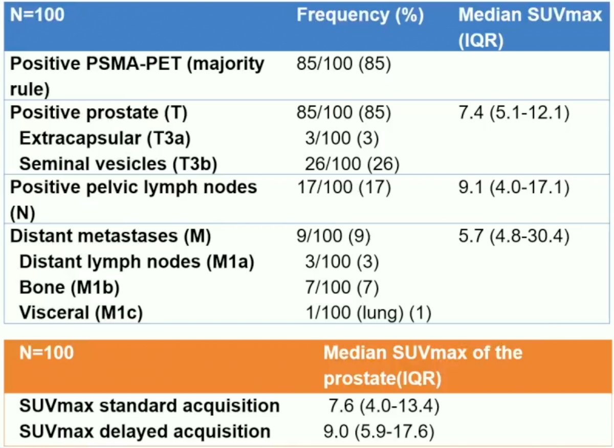
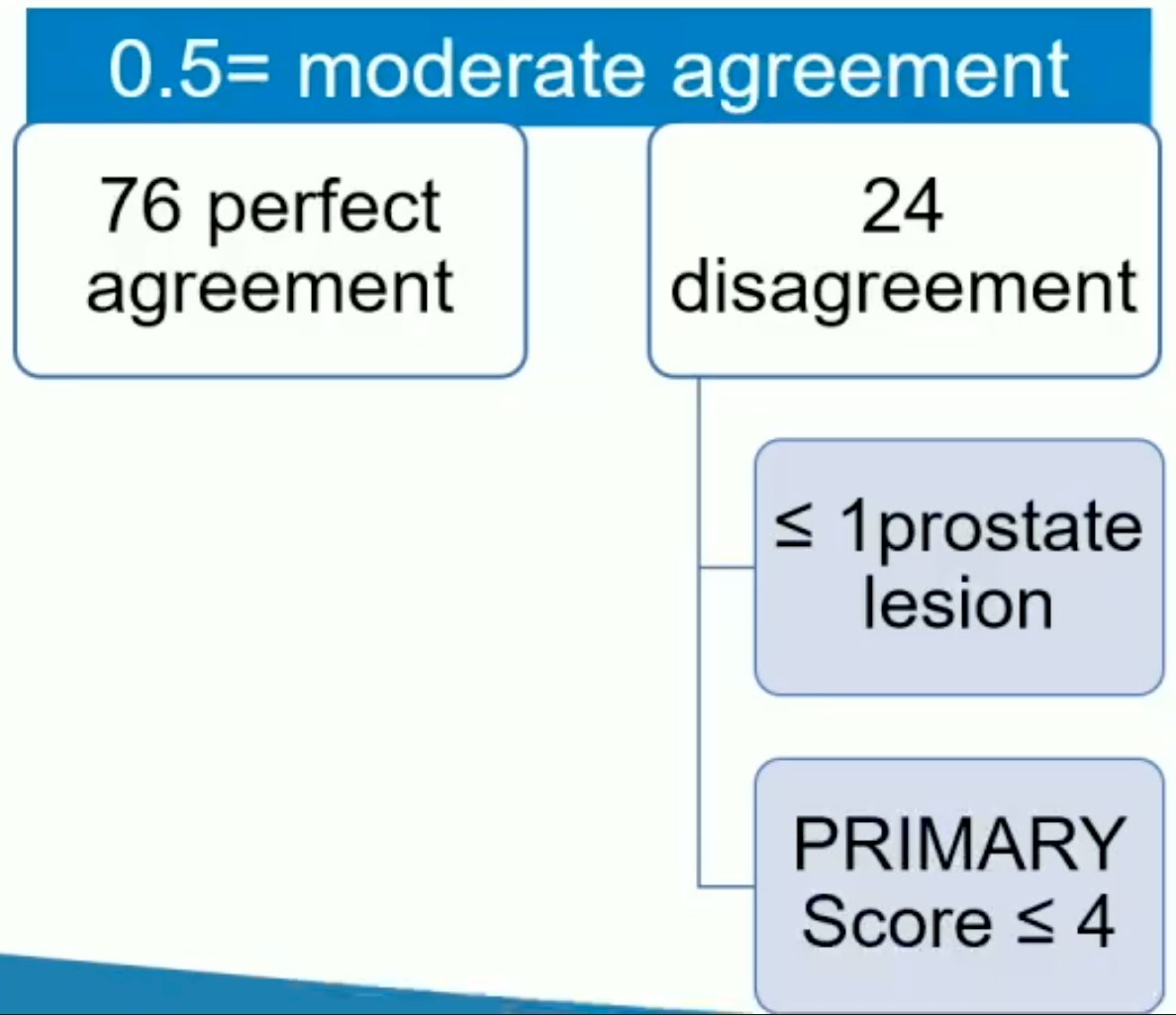
The inter-reader agreement was noted to be moderate (Kappa value= 0.50). Of the 100 cases, there was perfect agreement for 76% of the obtained images.
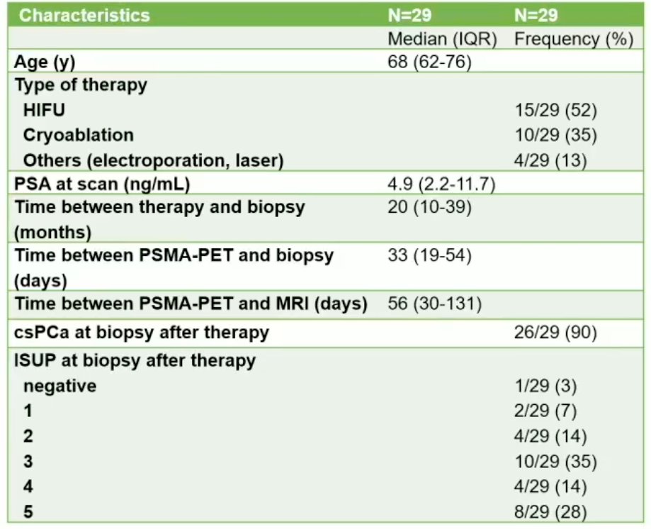
Next, the investigators focused on the subgroup of 29 patients with an available biopsy plus an MRI. 52% had undergone HIFU, 35% cryoablation, and 13% other focal therapy modalities. The median time between focal therapy and repeat biopsy was 20 months. The median time between the PSMA-PET/CT and the repeat biopsy and MRI were about one and 2 months, respectively. Of the 29 patients, 26 (90%) had csPCa detected on the post-treatment biopsy.
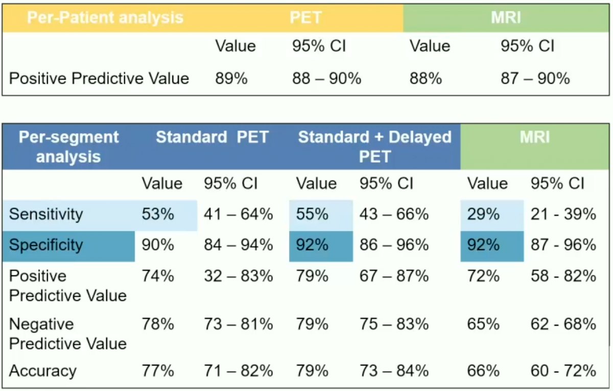
On a per-patient level, the positive predictive values for PSMA-PET/CT and MRI were comparable (89% and 88%, respectively). However, on a per-segment analysis level, the sensitivity of PET, whether using standard acquisition alone or standard + delayed acquisitions, was higher compared to MRI (53-55% versus 29%). The specificity levels were comparable (90-92% versus 92%).
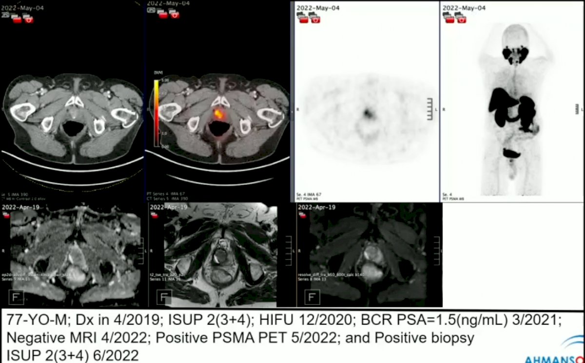
Dr. Jafarvand next highlighted a case of a 77-year-old patient with evidence of Grade Group 2 prostate cancer on repeat biopsy, but with discordant imaging results (PET+/MRI-), highlighting the benefit of PSMA-PET/CT in this setting.
Dr. Jafarvand concluded that these results suggest that PSMA-PET/CT has the potential to improve the localization of recurrent prostate cancer after focal therapy and help guide targeted biopsies for improved detection of persistent/recurrent csPCa following focal therapy . She suggested that further investigation with prospective studies within larger populations will be needed to further delineate the role of PSMA-PET/CT in the post-focal therapy setting.
Presented by: Mahbod Jafarvand, MD, Resident Physician, Department of Radiology and Nuclear Medicine, University of California, Los Angeles, CA
Written by: Rashid K. Sayyid, MD, MSc – Society of Urologic Oncology (SUO) Clinical Fellow at The University of Toronto, @rksayyid on Twitter during the 2023 Society of Nuclear Medicine and Molecular Imaging (SNMMI) Annual Meeting, Chicago, IL, Sat, June 24 – Tues, June 27, 2023.