Endoscopic Approaches and Emerging Novel Treatments for Upper Tract Urothelial Carcinoma

Upper tract urothelial carcinoma, comprising either the renal pelvis or ureter, is rarer than urothelial carcinoma of the bladder accounting for only 5-10% of all urothelial carcinomas1 (epidemiology of upper tract urothelial cancer, Wallis CJD). However, similar to bladder urothelial carcinoma, not all upper tract urothelial cancer is high-risk and/or patients may not be fit for radical surgery. Although a radical nephroureterectomy is the gold standard treatment for upper tract urothelial cancer, there has been an impetus for developing and assessing less aggressive, less invasive treatment modalities2-4. As such, this article will discuss endoscopic (ureteroscopic and percutaneous), kidney-sparing treatment modalities for upper tract urothelial cancer, as well as focus on several emerging treatments.

Indications for Kidney Sparing/Endoscopic Management

  • Imperative – patients with contraindications for radical surgery: (i) a solitary functioning kidney, (ii) bilateral upper tract urothelial cancer, (iii) baseline renal insufficiency, (iv) poor candidacy for hemodialysis or renal transplantation, (v) significant comorbidities
  • Elective – patients with low-risk non-muscle invasive upper tract urothelial cancer. May be eligible for a partial nephrectomy, segmental ureterectomy, or endoscopic management
The 2017 European Association of Urology Guidelines on upper tract urothelial cancer5 suggest that ureteroscopic endoscopic ablation can be considered in patients with clinically low-risk cancer in addition to the following situations: (i) a laser generator and equipment is available for biopsies, (ii) when a flexible rather than rigid ureteroscope is available, (iii) the patient is informed of the need for earlier or closer surveillance, and (iv) complete tumor resection is achievable. Furthermore, the Guidelines suggest that percutaneous ablation can be considered for low-risk upper tract urothelial cancer in the renal pelvis, particularly for tumors in the lower calyceal system that are inaccessible or difficult to manage by flexible ureteroscopy.

Ureteroscopic Management

Endoscopic evaluation, typically by ureteroscopy, is crucial for the initial diagnosis, risk stratification, and subsequent treatment planning for patients with upper tract urothelial cancer. Information from endoscopy helps assess tumor location, multifocality, architecture and allows the clinician to obtain a tissue diagnosis. However, ureteroscopic biopsies can be technically challenging given the limited ureteroscopic biopsy instruments that can traverse the small working channel. Some experts have advocated that the visual characteristics of the tumor may be able to predict disease aggressiveness, such as sessile-appearing tumors being more likely to be higher grade/stage .6 Several techniques for achieving a tissue diagnosis have been described, including multiple urine and washing samples and multiple quality biopsies to ensure sufficient tissue for pathological assessment; the most commonly used biopsy tools include the Piranha forceps (Boston Scientific, Marlborough, MA) and the BIGopsy forceps (Cook Medical, Bloomington, IN).2 For larger, papillary tumors, a stone basket (ie. nitinol) can be used for snaring and debulking the tumor.  

One of the benefits of a ureteroscopic approach is that a single procedure can be both diagnostic and therapeutic. When possible, use of a ureteral access sheath is ideal in that it allows atraumatic multiple insertions of the ureteroscope, especially important when encountered with a large tumor volume7. Following multiple biopsies for tissue diagnosis, a laser can be used both for additional tumor resection and fulguration (for hemostasis) of the tumor bed. The most commonly used lasers are the holmium:yttrium-aluminum-garnet (YAG) laser and the neodymium-doped (Nd):YAG laser. The holmium laser is typically better suited for smaller tumors, however, it requires contact with the tissue in order to be effective. The Nd: YAG laser has a smaller wavelength, is able to penetrate deeper (5-6 mm), and does not require direct tissue contact. Although less readily available, electrocautery resection is also possible with a 10-13Fr rigid ureteral resectoscope (Karl Storz Endoscopy, Tuttlingen, Germany); this scope allows resection of tumors similar to a loop used for transurethral resection of bladder tumors2.

Currently, there is no Level I evidence assessing oncologic outcomes of patients undergoing ureteroscopic management of upper tract urothelial cancer. Retrospective series to date are all limited to fewer than 100 patients. The largest retrospective series with more than two years of follow-up was published by Grasso and colleagues (n=82)8 noting 81% recurrence rate in the upper tract, 57% in the bladder, 19% of patients progressing to surgical resection, and a 74% and 87% overall and cancer-specific survival rate, respectively. Petros et al summarized these retrospective studies with the following outcomes:2

  • Median follow-up: 24-58 months
  • Upper tract recurrence rate: 65%
  • Bladder recurrence rate: 44%
  • Progression to surgical resection rate: 0-33%
  • Overall survival rate: 35-100%
  • Cancer-specific survival rate: 70-100%
In general, ureteroscopic management of upper tract urothelial cancer is associated with high risk of recurrence and a not insignificant rate of progression to more radical surgery.

Percutaneous Management

Antegrade percutaneous endoscopic treatment of upper tract urothelial carcinoma is typically reserved for patients with low-grade, large volume tumors that are either anatomically or technically challenging for ureteroscopic management, ie. lower pole tumors. This approach is particularly advantageous for patients that have had a prior cystectomy and urinary diversion. A major benefit of percutaneous management is the ability to use larger instruments that are able to fit through a nephroscope, including loop cautery for debulking large tumors. All of the laser biopsy instruments listed above for ureteroscopic management are also feasible for the percutaneous approach. The primary risk of percutaneous management of upper tract urothelial carcinoma (aside from those traditionally associated with percutaneous management of nephrolithiasis) is disruption of the urothelium, which may lead to a theoretically increased risk of tumor seeding into the retroperitoneum.2

Similar to ureteroscopic management of upper tract urothelial carcinoma, oncologic outcomes have relied on retrospective studies of fewer than 100 patients. However, one study by Motamedinia et al.9 identified 141 patients who underwent percutaneous resection with a median follow-up of 66 months. They noted that recurrence occurred in 37% of low-grade patients and 63% of high-grade patients, with a median time to recurrence of 71.4 vs 36.4 months, respectively. On multivariable analysis, grade was the only predictor of recurrence (HR 2.12, p = 0.018) and radical nephroureterectomy was avoided in 87% of patients. Petros and colleagues2 summarized oncologic outcomes of percutaneous management among studies with a minimum 2-year follow-up (n=361):

  • Median follow-up: 24-66 months
  • Upper tract recurrence rate: 40%
  • Bladder recurrence rate: 24%
  • Progression to surgical resection rate: 6-50%
  • Overall survival rate: 40-90%
  • Cancer-specific survival rate: 75-100%

Emerging Novel Treatments

Several new and exciting treatments are under development with regards to minimally invasive treatment of upper tract urothelial cancer. As highlighted above, the recurrence rate for endoscopic management is high (40-65% on pooled analysis)2 and these approaches are not well suited to treatment of multifocal disease or carcinoma in situ. Thus, much research has been directed for improving the deliverability of topical agents to the upper tracts. One of the challenges is difficulty concentrating therapeutic levels of these agents in the upper tract for more than a brief period of time secondary to ureteral peristalsis rapidly draining topical treatment from the pelvis and ureter. Specifically, mitomycin C exposure time to the urothelium is critical for efficacy.10 RTGel™ is a reverse-thermal hydrogel composed of a combination of polymers that allows it to exist as a liquid at cold temperatures but solidifies to a gel state at body temperature.11 This product was developed to address the constraints of the upper urinary tract, where continuous urine production and ureteral peristalsis prevents drug retention in the upper tract. Subsequently, MitoGel Ôwas developed as a novel formulation of RTGel combined with mitomycin C. The hypothesis for MitoGel is that upon delivery to the upper urinary tract, MitoGel would gelatinize and urine would produce a slow dissolution of the gel, allowing a sustained release of mitomycin C into the upper tract allowing prolonged exposure to the urothelium. Using a preclinical swine animal model (n=23), Donin et al,11 noted that after antegrade instillation of MitoGel, the product remained visible in the pelvicalyceal system on fluoroscopic and computed tomography imaging for 4-6 hours after instillation. Furthermore, on necropsy, they noted that mitomycin C plasma levels were well within acceptable safety thresholds and that there was no evidence of urinary obstruction, acute kidney injury, sepsis, or myelosuppression. Donin and colleagues subsequently confirmed these safety results in a study assessing six once-weekly unilateral retrograde instillations of Mitogel.12

The OLYMPUS study (NCT02793128) is a prospective single-arm ongoing clinical trial designed to assess the efficacy, safety, and tolerability of MitoGel in patients with low grade, noninvasive upper tract urothelial cancer. Eligible patients are treated with MitoGel once weekly for a total of six times in a retrograde fashion; patients demonstrating a complete response are treated with MitoGel once monthly for a total of 11 instillations as maintenance, or until the first recurrence. The primary outcomes are complete response rate defined as the percent of patients with complete response at the primary disease evaluation visit (~11 weeks), and adverse event rates (over ~2 years). Secondary outcomes include:

(i) Long-term durability of complete response (12 months)
(ii) Complete response rates at 3, 6, and 9 months
(iii) Partial response to treatment (~11 weeks)
(iv) Mitomycin C level in blood plasma

The target recruitment goal is 71 patients with an estimated study completion date of February 2020.

Previous studies assessing differential gene expression between upper tract and bladder urothelial carcinoma using microarray data suggest that upper tract tumors tend to have high expression of genes associated with a luminal subtype.13 Furthermore, one particular gene highly expressed in upper tract tumors is SLITRK6, an integral membrane protein known to have high levels of expression in certain carcinomas, but low levels in the majority of other tissues.14 An antibody to SLITRK6 protein has been developed and linked to a cytotoxic agent called monomethyl auristatin E (AGS15E), a microtubule-disrupting agent.15, 16 ASG-15C was chosen among seven anti-SLITRK6 antibodies and monomethyl auristatin E was chosen due to its efficacy in tumor inhibition and regression. Upon binding to SLITRK6, ASG-15ME is rapidly internalized and trafficked to lysosomes and early endosomes. An ongoing Phase I dose escalation trial of AGS15E in patients with metastatic urothelial carcinoma presented initial interim trial results at the ESMO 2016.17 This trial includes patients previously treated with ≥ 1 prior chemotherapy regimens; SLITRK6 expression was determined by immunohistochemistry. Disease assessments were performed every 8 weeks using RECIST v 1.1 criteria and ASG-15ME was administered IV weekly for 3 out of every 4 weeks until no further benefit. Six dose levels were assessed: 0.1, 0.25, 0.5, 0.75, 1, and 1.25 mg/kg. At the time of analysis, 51 pts were enrolled and 93% were SLITRK6 positive. Of 42 evaluable patients at doses considered active (doses ≥0.5 mg/kg), one had a complete response and 13 had a partial response (ORR =33%). Adverse event rate was high (91%) and the most common treatment-related adverse event was fatigue (44%). Serum concentrations of ASG-15ME decreased multi-exponentially and half-life was 3.1 days. In heavily pre-treated individuals, novel antibody-drug conjugates continue to be explored for therapeutic benefit.

Conclusions  

Endoscopic management of upper tract urothelial cancer is technically feasible but associated with high rates of recurrence and non-insignificant rates of progression necessitating radical surgical treatment. Endoscopic management should be reserved for low-grade tumors and patients that have contraindications to radical nephroureterectomy. Research is ongoing, particularly for improving delivery of topical agents to decrease recurrence rates.

Published Date: April 15th, 2019
Written by: Zachary Klaassen, MD, MSc
References:
  1. Siegel RL, Miller KD, Jemal A. Cancer statistics, 2019. CA Cancer J Clin. 2019;69:7-34.
  2. Petros FG, Li R, Matin SF. Endoscopic Approaches to Upper Tract Urothelial Carcinoma. Urol Clin North Am. 2018;45:267-86.
  3. Samson P, Smith AD, Hoenig D, Okeke Z. Endoscopic Management of Upper Urinary Tract Urothelial Carcinoma. J Endourol. 2018;32:S10-S6.
  4. Cutress ML, Stewart GD, Zakikhani P, Phipps S, Thomas BG, Tolley DA. Ureteroscopic and percutaneous management of upper tract urothelial carcinoma (UTUC): systematic review. BJU Int. 2012;110:614-28.
  5. Roupret M, Babjuk M, Comperat E, Zigeuner R, Sylvester RJ, Burger M, et al. European Association of Urology Guidelines on Upper Urinary Tract Urothelial Carcinoma: 2017 Update. Eur Urol. 2018;73:111-22.
  6. Williams SK, Denton KJ, Minervini A, Oxley J, Khastigir J, Timoney AG, et al. Correlation of upper-tract cytology, retrograde pyelography, ureteroscopic appearance, and ureteroscopic biopsy with histologic examination of upper-tract transitional cell carcinoma. J Endourol. 2008;22:71-6.
  7. Raman JD, Park R. Endoscopic management of upper-tract urothelial carcinoma. Expert Rev Anticancer Ther. 2017;17:545-54.
  8. Grasso M, Fishman AI, Cohen J, Alexander B. Ureteroscopic and extirpative treatment of upper urinary tract urothelial carcinoma: a 15-year comprehensive review of 160 consecutive patients. BJU Int. 2012;110:1618-26.
  9. Motamedinia P, Keheila M, Leavitt DA, Rastinehad AR, Okeke Z, Smith AD. The Expanded Use of Percutaneous Resection for Upper Tract Urothelial Carcinoma: A 30-Year Comprehensive Experience. J Endourol. 2016;30:262-7.
  10. De Bruijn EA, Sleeboom HP, van Helsdingen PJ, van Oosterom AT, Tjaden UR, Maes RA. Pharmacodynamics and pharmacokinetics of intravesical mitomycin C upon different dwelling times. Int J Cancer. 1992;51:359-64.
  11. Donin NM, Duarte S, Lenis AT, Caliliw R, Torres C, Smithson A, et al. Sustained-release Formulation of Mitomycin C to the Upper Urinary Tract Using a Thermosensitive Polymer: A Preclinical Study. Urology. 2017;99:270-7.
  12. Donin NM, Strauss-Ayali D, Agmon-Gerstein Y, Malchi N, Lenis AT, Holden S, et al. Serial retrograde instillations of sustained release formulation of mitomycin C to the upper urinary tract of the Yorkshire swine using a thermosensitive polymer: Safety and feasibility. Urol Oncol. 2017;35:272-8.
  13. Sanford T, Porten S, Meng MV. Molecular Analysis of Upper Tract and Bladder Urothelial Carcinoma: Results from a Microarray Comparison. PLoS One. 2015;10:e0137141.
  14. Aruga J, Yokota N, Mikoshiba K. Human SLITRK family genes: genomic organization and expression profiling in normal brain and brain tumor tissue. Gene. 2003;315:87-94.
  15. Vlachostergios PJ, Jakubowski CD, Niaz MJ, Lee A, Thomas C, Hackett AL, et al. Antibody-Drug Conjugates in Bladder Cancer. Bladder Cancer. 2018;4:247-59.
  16. Morrison K, Challita-Eid PM, Raitano A, An Z, Yang P, Abad JD, et al. Development of ASG-15ME, a Novel Antibody-Drug Conjugate Targeting SLITRK6, a New Urothelial Cancer Biomarker. Mol Cancer Ther. 2016;15:1301-10.
  17. Petrylak D, Heath E, Sonpavde G, George S, Morgans AK, Eigl BJ. Interim analysis of phase 1 dose escalation trial of the antibody-drug conjugate (ADC) ASG15E (ASG15ME) in patients (Pts) with metastatic urothelial cancer (mUC). Ann Oncol. 2016;27:266-95.

Nonsurgical Focal Therapy for Renal Tumors

As has been highlighted in the accompanying article on the Epidemiology and Etiology of Kidney Cancer, cancers of the kidney and renal pelvis comprise the 6th most common newly diagnosed tumors in men and 10th most common in women.1 With the increasing use of abdominal imaging, a growing number of small renal masses are being detected. In fact, 13 to 27% of abdominal imaging studies demonstrate incidental renal lesions unrelated to the reason for the study2 and approximately 80% of these masses are malignant.3Thus, a large number of small, incidentally-detected renal masses are now being diagnosed. Due to the increase in diagnosis of small renal masses and the general predilection for diagnosis of renal tumors in older adults (typically diagnosed between age 50 and 70 years), the paradigm for treatment of renal tumors has focused on minimally invasive approaches and nephron sparing in the past few years.

According to the American Urological Association guidelines on the management of stage 1 renal tumors, nephron sparing surgery (partial nephrectomy) is recommended.4 However, renal mass ablation is considered an alternative, particularly among the elderly and comorbid.4 Renal ablation may be undertaken by percutaneous approaches (nonsurgical) or through laparoscopic or open approaches.

Rationale for Focal Therapy

As with any tumor site, focal ablative therapies offer several potential advantages to traditional surgical approaches. First, focal ablative therapies are less physiologically demanding on the patient than extirpative surgery. As a result, these may often be performed as ambulatory day surgical procedures with a much shorter convalescence and fewer complications when compared to laparoscopic partial nephrectomy.5 Second, renal mass ablation is associated with comparable post-operative renal function when compared to partial nephrectomy.5,6 Third, while laparoscopic partial nephrectomy is a technically challenging operation, requiring advanced laparoscopic skills for tumour resection and renal reconstruction,7 focal ablation (either via laparoscopic or percutaneous approach) allows minimally-invasive treatment of renal tumors with relative technical simplicity.5 Finally, renal mass ablation may be accomplished by a variety of approaches including open, laparoscopic, and percutaneous approaches.

While long-term data are lacking, intermediate term data (with a median follow-up of approximately 3.5 years) suggest that cancer control is similar between renal tumor ablation (using laparoscopic cryotherapy) and minimally-invasive partial nephrectomy.6

Indications for Focal Therapy of Renal Tumors

Treatment choice in the management of small renal masses depends on a complex interplay of patient preference, tumor characteristics, host (patient) factors including age and comorbidity, and the expertise/ability of the treating physician. A number of indications have been well-recognized for the use of renal tumor ablation. Ablation is indicated for patients with small renal tumors who are: poor surgical candidates or at high risk of renal insufficiency. Patients may be at risk of renal insufficiency due to underlying nephron-threatening conditions such as diabetes or hypertension, due to a solitary kidney (either congenital or due to prior nephrectomy), or due to oncologic factors such as bilateral tumors or hereditary syndromes which predispose to recurrent, multifocal tumors.

However, given the good outcomes of renal mass ablation in the treatment of small renal masses among these patients, a number of authors have now advocated the use of renal mass ablation in otherwise healthy patients.8

Approaches to Focal Therapy

Non-surgical focal therapy refers to a therapeutic strategy, rather than a specific treatment modality. A number of different focal therapy modalities have been employed in the treatment of small renal masses. Foremost among these are cryoablation and radiofrequency ablation (RFA).

Prior to ablation, the American Urologic Association guidelines recommend biopsy of the renal mass either prior to ablation or at the time of treatment.9

Cryotherapy

Cryoablation, also known as cryotherapy, is the therapeutic use of extremely cold temperature. While first employed in the treatment of breast, cervical, and skin cancers, cryoablation has subsequently been used in the treatment of a variety of benign and malignant conditions. Initially, liquified air was used, then solidified carbon dioxide, liquid oxygen, liquid nitrogen, and finally argon gas. Today, the majority of commercially available systems rely on argon gas.

It wasn’t until Onik et al. identified that the cryogenic ice-tissue interface was highly echogenic on ultrasound that an accurate, controlled treatment of intra-abdominal malignancies could be undertaken.10 Today, cryotherapy of renal tumors is undertaken under real-time imaging.

Ablation during cryoablation occurs during both the freezing and thawing phases of the treatment cycle. During freezing, the rapid decrease in temperature immediately adjacent to the probe causes the formation of intracellular ice crystals which lead to mechanical trauma to plasma membranes and organelles and subsequent cell death through ischemia and apoptosis.11 More distal to the probe, a slower freezing process occurs in which extracellular ice crystals form, causing depletion of extracellular water and inducing an osmotic gradient which causes intracellular dehydration. During the thaw cycle, extracellular ice crystals melt leading to an influx of water back into the cells, resulting in cellular edema. In addition to these cellular effects, the freezing cycle results in injury to the blood vessel endothelium resulting in platelet activation, vascular thrombosis and tissue ischemia. The result of these process is coagulative necrosis, cellular apoptosis, fibrosis and scar formation. Due to evidence that multiple freeze-thaw cycles led to larger areas of necrosis, the current treatment paradox suggests a double freeze-thaw cycle.

For optimal cellular death, the preferred target temperature for cryotherapy is at or below -40o C. As temperatures at the edge of the ice ball are 0o C, most authors suggest that the ice ball extends at least 5 or 10mm beyond the edge of the target lesion. In some cases, this will require the use of multiple probes.

Radiofrequency Ablation

In contrast to cryotherapy which utilizes freeze-thaw cycles to induce cellular damage, radiofrequency ablation (RFA) relies upon radiofrequency energy to heat tissue until cellular death. Using monopolar alternating electrical current at a frequency of 450 to 1200 kHz, RFA induces vibrations of ions within the tissue which leads to molecular friction and heat production. The resulting increased intracellular temperature leads to cellular protein denaturation and cell membrane disintegration. The success of RFA treatment depends on the power delivered, the resulting maximal temperature achieved, and the duration of ablation.

A number of variations in RFA delivery have been described: temperature- or impedance-based guidance, single or multiple tines, “wet” vs “dry” ablation, and mono- or bi-polar electrodes.

Unlike cryoablation which relies upon real-time imaging guidance, RFA may be guided by either temperature-based or impedance-based monitoring. Systems which rely on temperature-based guidance measure temperature at the tip of the electrode. However, they do not measure temperature within the surrounding tissue. Systems which rely on impedance-based guidance measure the resistance to alternating current (the impedance). These systems are calibrated to achieve a predetermined impedance level. There is no data to support the superiority of either of these approaches. For temperature-based systems, the target is 105o C with a minimum of 70o C during the heating cycle. For impedance-based systems, the target is 200 to 500 ohms, which is achieved by progressively increasing the power beginning from 40-80W to 130-200W at a rate of 10W/minute.

A number of studies have demonstrated that multi-tine electrodes are associated with more complete tissue necrosis and improved treatment outcomes.12

In addition to the guidance approach and number of tines, RFA technology may be stratified according to “wet” vs. “dry” approach. Through the tissue ablation process, tissue desiccation leads to charring which can increase impedance. This in turn increases the resistance to the current emanating from the electrode and limits the size of the ablation field. A “dry” approach functions within these limitations and cannot treat more than 4cm with a single electrode. In contrast, a “wet” approach continuously infuses saline through the probe tip. This cools the tissue and prevents the tissue charring. As a result, larger ablation zones are possible.

Finally, energy delivery may be either through monopolar or bipolar electrodes. The benefit of bipolar electrodes is both increased temperature generation13 and a larger treatment field.14

The efficacy of RFA is affected not only by the characteristics of the tissue being treated but also by the surrounding tissues. For example, large vessels may dissipate heat and result in relative undertreatment of adjacent tissues.

Monitoring following Focal Therapy

The definition of treatment success following renal mass focal ablation has been controversial. Currently, radiographic assessment utilizing computed tomography or magnetic resonance imaging is considered an accepted measure of treatment effect.15 Typically, this is performed 4-12 weeks following treatment. However, some rely on post-ablation biopsy to confirm treatment success though this is not well accepted.

The most reliable radiographic marker of successful ablation is the lack of contrast enhancement, corresponding to complete tissue destruction.16 Persistent enhancement is considered incomplete treatment and re-treatment or an alternative treatment strategy may be warranted. Alternatively, subsequent enhancement on surveillance imaging in an area with prior loss of enhancement suggests local recurrence.17 Many tumors following cryoablation have a significant reduction in tumor size while this is uncommon following RFA.

The AUA guidelines recommend contrast enhanced CT or MUI at 3 and 6 months following treatment and then each year for the following 5 years.9

Oncologic Outcomes

Long-term outcomes are lacking for renal ablation techniques. The summary data from the AUA guidelines panel suggests local recurrence free rates of approximately 90% for patients undergoing cryoablation and 87% for patients undergoing RFA.4 Outcomes between cryoablation and RFA appear to be comparable. Compared to partial nephrectomy, the available data suggest higher rates of local recurrence despite shorter follow-up. However, metastasis-free survival and cancer-specific survival appear to be comparable.

Complications

Major complications following renal mass ablation are uncommon. Further, percutaneous, nonsurgical ablation has lower complication rates than other approaches.18 As with oncologic outcomes, complication rates are comparable between RFA and cryoablation. Major urologic complications occurred in 3.3-8.2% of patients undergoing ablation while non-urologic complications occurred in 3.2-7.2%. These rates are lower than extirpative approaches including open or laparoscopic nephrectomy.

The most common complication is pain or paresthesia at the percutaneous access site.19 The most concerning complications relate to inadvertent injury to intra-abdominal organs. A variety of tumor characteristics including anterior location, proximity to collecting system and those without easy percutaneous access increase the risk of complications when percutaneous ablation is undertaken. Permanent urologic damage including injury to calyces, the ureteropelvic junction, or the ureter is uncommon.20

Hemorrhage is the most common serious complication of cryoablation. This is less common with RFA. Bleeding is more common when multiple probes are used to treat large tumors.21

Published Date: November 20th, 2018

References:

1. Siegel RL, Miller KD, Jemal A. Cancer statistics, 2018. CA: a cancer journal for clinicians 2018;68:7-30.

2. Gill IS, Aron M, Gervais DA, Jewett MA. Clinical practice. Small renal mass. The New England journal of medicine 2010;362:624-34.

3. Frank I, Blute ML, Cheville JC, Lohse CM, Weaver AL, Zincke H. Solid renal tumors: an analysis of pathological features related to tumor size. The Journal of urology 2003;170:2217-20.

4. Campbell SC, Novick AC, Belldegrun A, et al. Guideline for management of the clinical T1 renal mass. The Journal of urology 2009;182:1271-9.

5. Desai MM, Aron M, Gill IS. Laparoscopic partial nephrectomy versus laparoscopic cryoablation for the small renal tumor. Urology 2005;66:23-8.

6. Fossati N, Larcher A, Gadda GM, et al. Minimally Invasive Partial Nephrectomy Versus Laparoscopic Cryoablation for Patients Newly Diagnosed with a Single Small Renal Mass. Eur Urol Focus 2015;1:66-72.

7. Aboumarzouk OM, Stein RJ, Eyraud R, et al. Robotic Versus Laparoscopic Partial Nephrectomy: A Systematic Review and Meta-Analysis. European Urology 2012;62:1023-33.

8. Stern JM, Gupta A, Raman JD, et al. Radiofrequency ablation of small renal cortical tumours in healthy adults: renal function preservation and intermediate oncological outcome. BJU international 2009;104:786-9.

9. Donat SM, Diaz M, Bishoff JT, et al. Follow-up for Clinically Localized Renal Neoplasms: AUA Guideline. The Journal of urology 2013;190:407-16.

10. Onik G, Gilbert J, Hoddick W, et al. Sonographic monitoring of hepatic cryosurgery in an experimental animal model. AJR Am J Roentgenol 1985;144:1043-7.

11. Baust JG, Gage AA. The molecular basis of cryosurgery. BJU international 2005;95:1187-91.

12. Rehman J, Landman J, Lee D, et al. Needle-based ablation of renal parenchyma using microwave, cryoablation, impedance- and temperature-based monopolar and bipolar radiofrequency, and liquid and gel chemoablation: laboratory studies and review of the literature. J Endourol 2004;18:83-104.

13. Nakada SY, Jerde TJ, Warner TF, et al. Bipolar radiofrequency ablation of the kidney: comparison with monopolar radiofrequency ablation. J Endourol 2003;17:927-33.

14. McGahan JP, Gu WZ, Brock JM, Tesluk H, Jones CD. Hepatic ablation using bipolar radiofrequency electrocautery. Acad Radiol 1996;3:418-22.

15. Matin SF, Ahrar K, Cadeddu JA, et al. Residual and recurrent disease following renal energy ablative therapy: a multi-institutional study. The Journal of urology 2006;176:1973-7.

16. Matsumoto ED, Watumull L, Johnson DB, et al. The radiographic evolution of radio frequency ablated renal tumors. The Journal of urology 2004;172:45-8.

17. Matin SF. Determining failure after renal ablative therapy for renal cell carcinoma: false-negative and false-positive imaging findings. Urology 2010;75:1254-7.

18. Johnson DB, Solomon SB, Su LM, et al. Defining the complications of cryoablation and radio frequency ablation of small renal tumors: a multi-institutional review. The Journal of urology 2004;172:874-7.

19. Farrell MA, Charboneau WJ, DiMarco DS, et al. Imaging-guided radiofrequency ablation of solid renal tumors. AJR Am J Roentgenol 2003;180:1509-13.

20. Johnson DB, Saboorian MH, Duchene DA, Ogan K, Cadeddu JA. Nephrectomy after radiofrequency ablation-induced ureteropelvic junction obstruction: potential complication and long-term assessment of ablation adequacy. Urology 2003;62:351-2.

21. Lehman DS, Hruby GW, Phillips CK, McKiernan JM, Benson MC, Landman J. First Prize (tie): Laparoscopic renal cryoablation: efficacy and complications for larger renal masses. J Endourol 2008;22:1123-7.

First Line Therapy of Metastatic Clear Cell RCC

Background

Kidney cancer represents 5% of all new cancer diagnoses in the United States, with approximately 64,000 new cases and 14,970 deaths in 2018.1,2 The most common type of kidney cancer is renal cell carcinoma (RCC) and the most common histologic subtype of RCC is clear cell RCC, accounting for over 80% of cases.3 RCC is more common in men than women and typically occurs in the sixth to eighth decade of life.1 Localized kidney cancer can often be cured with definitive surgery, with 5-year survival reaching over 90%.However, for patients with advanced disease, 5-year survival remains poor at 11.7% and much progress is needed to develop novel therapies for advanced RCC.4

Risk Stratification

The current treatment paradigm for metastatic clear cell RCC requires stratification of patients into favorable, intermediate, or poor risk disease. Several validated models for risk stratification exist, including the International Metastatic Renal Cell Carcinoma Database Consortium model (IMDC)5 and Memorial Sloan Kettering Cancer Center model (MSKCC).6 Both criteria include time from diagnosis to systemic treatment of less than one year, performance status, as well as hemoglobin and calcium (Table 1). The main differences between these models are that the MSKCC model includes LDH and the IMDC includes neutrophil and platelet count as unique risk factors. Patients with no risk factors fall into the favorable risk group, patients with one to two risk factors are in the intermediate risk group, and those with three or more risk factors are in the high-risk group for both prognostic models. Contemporary clinical trials have found that drug effectiveness may vary depending on risk stratification which has led the FDA to approve some therapies for only certain risk groups.7 Thus, risk stratification is important for the clinician, not only for discussing prognosis with patients, but also for treatment selection. 
table-1-first-line-therapy-clear-cell-RCC@2x.jpg

Favorable Risk Patients

For patients with no adverse risk factors, sunitinib and pazopanib are the preferred first line treatment options, recommended by both the National Comprehensive Cancer Network (NCCN) as well as the European Association of Urology (EAU).8,9 Sunitinib is an oral multikinase inhibitor which targets vascular endothelial growth factor receptors (VEGFR) and platelet-derived growth factor receptors (PDGFR). A phase III trial comparing sunitinib to interferon-α showed that sunitinib significantly improved median progression free survival (11 months vs 5 months) and had a higher overall response rate as well when compared with interferon alfa (31% vs 6%).10 Sunitinib also demonstrated longer overall survival in a follow up study, 26.4 months vs 21.8 months.11 Severe adverse events (grade 3-4 toxicities) were minimal, and the most common adverse events were diarrhea, fatigue, and nausea. Hypertension was one notable side effect of sunitinib, which was not seen with interferon alfa. Based on the data above, sunitinib was granted FDA approval in 2007 and has been the benchmark for many future clinical trials in the mRCC space. 

Pazopanib is another oral multikinase inhibitor, which targets VEGFR-1,2,3, PDGFR- and , and c-KIT.12 Pazopanib was established as a safe and efficacious therapy for mRCC based on a large randomized, placebo controlled trial in patients who were treatment naive or cytokine pretreated.12 In this trial of 435 patients, pazopanib increased progression-free survival (9.2 months vs 4.2 months) compared with placebo, both in the treatment naïve cohort as well as the cytokine pretreated cohort. The objective response rate to pazopanib was 30% and pazopanib was well tolerated. Unique toxicities of pazopanib included notable grade 3 hepatotoxicity  30% of patients had elevated ALT and 21% had elevated AST. These results granted pazopanib FDA approval in October 2009. A subsequent phase III study (COMPARZ) with 1,110 patients compared sunitinib to pazopanib, and pazopanib was found to be noninferior to sunitinib with respect to progression-free survival and overall survival.13 Median overall survival was 28.4 months in the pazopanib arm compared with 29.3 months in the sunitinib arm. In terms of safety, similar percentages of patients in both sunitinib and pazopanib experienced dose interruptions of one week or greater, 44% and 49% respectively. Patients in the pazopanib arm more frequently discontinued therapy based on abnormal liver function tests. Patients taking sunitinib had a higher risk of abnormal hematologic labs including leukopenia, thrombocytopenia, neutropenia, and anemia. 

Additional FDA approved therapies for first line management of favorable risk patients include high dose interleukin 2 (HD IL-2)14-17, interferon plus bevacizumab18, and sorafenib.19 These therapies are less commonly used given their tolerability and toxicity profiles. HD IL-2 deserves special mention here given its ability to induce complete responses in a small subset of patients. A recent abstract describing overall survival from the PROCLAIM database show that of favorable risk patients, median overall survival is 63.3 months and 2 year overall survival is 77.6%.14 Of course, patients who are given HD IL-2 are a very carefully selected robust cohort, and cross-trial comparisons are challenging to make. 

Intermediate and Poor Risk

Per the IMDC and MSKCC prognostic models, patients with one or two risk factors are classified as intermediate risk, and poor risk if they have three or more risk factors. Currently, the two major newcomers in this space are cabozantinib and the combination of ipilimumab and nivolumab.  Cabozantinib is a multikinase inhibitor of VEGFR, MET, and AXL.20 In the phase II CABOSUN study, 157 intermediate or poor risk patients were randomized to cabozantinib or sunitinib. In this population, cabozantinib increased median progression free survival (8.2 months vs 5.6 months) and improved overall response rate (33% vs to 12%) compared with sunitinib. Both sunitinib and cabozantinib had about a 67% grade 3/4 adverse event rate and had a similar toxicity profile, including fatigue, hypertension, and diarrhea. Sunitinib had a lower incidence of hand foot syndrome and weight loss compared with cabozantinib, but higher rates of neutropenia and thrombocytopenia. Given this data, Cabozantinib obtained FDA approval for the front-line treatment of mRCC in December 2017. 

The newest therapy to obtain FDA approval is the combination checkpoint inhibitor duo Ipilimumab and Nivolumab (Ipi/Nivo); it was approved in April of 2018, based on the results of CheckMate214.7 CheckMate 214 was a randomized, open-label trial comparing sunitinib with Ipi/Nivo. 1096 patients were enrolled, of which 847 were intermediate or poor risk. This study had a coprimary endpoint of overall survival, objective response rate, and progression free survival among patients who were intermediate or poor risk. The overall response rate was 42% with ipi/nivo vs 27% with sunitinib, with a complete response rate of 9% vs 1%. Median overall survival has not been reached with ipi/nivo vs 26 months for sunitinib. Progression free survival was 11.6 months for ipi/nivo compared with 8.4 months for sunitinib. 46% of patients receiving ipi/nivo experienced grade 3/4 toxicities compared with 63% of patients receiving sunitinib. The most common grade 3/4 adverse events with ipi/nivo was fatigue, diarrhea, and elevated lipase compared with hypertension, hand-foot syndrome, and increased lipase with sunitinib. 35% of patients required high dose corticosteroids for immune related toxicities with ipi/nivo.  With this data, the European Association of Urology has recommended that ipi/nivo be the new standard of care for patients with intermediate and poor risk disease, and the NCCN has also listed ipi/nivo as a category 1, preferred treatment option for patients with intermediate and poor risk disease.8,9
table-2-first-line-therapy-clear-cell-RCC@2x.png

Future Therapies

Front-line treatment options for mRCC are rapidly evolving.24 Data shown at ASCO and GU ASCO has demonstrated that antiangiogenic agents in combination with checkpoint inhibitors may prolong progression-free survival when compared with kinase inhibitors.25 Several phase III trials exploring this hypothesis are now underway (Table 3). IMmotion 151 is a randomized phase III trial comparing the combination of atezolizumab + bevacizumab vs sunitinib. Progression-free survival was 11.2 months in the intention to treat analysis for patients atezolizumab and bevacizumab, compared with 8.4 months in the sunitinib arm. JAVELIN Renal 100 is investigating avelumab in combination with axitinib and phase I results show a promising overall response rate of 54.5% out of 55 patients.26 KEYNOTE-426 is investigating pembrolizumab in combination with axitinib, compared with sunitinib alone.27 These trials are important and exciting for our patients with mRCC and their future results may alter the standard of care for frontline mRCC.
table-3-first-line-therapy-clear-cell-RCC@2x.png


Published Date: January 29th, 2019

Written by: Jason Zhu, MD
References:
  1. Siegel RL, Miller KD, Jemal A. Cancer statistics, 2018. CA: A Cancer Journal for Clinicians 2018;68:7-30.2018. at https://www.cancer.org/cancer/kidney-cancer/about/key-statistics.html.)
  2. Moch H, Gasser T, Amin MB, Torhorst J, Sauter G, Mihatsch MJ. Prognostic utility of the recently recommended histologic classification and revised TNM staging system of renal cell carcinoma. Cancer 2000;89:604-14.
  3. Motzer RJ, Jonasch E, Agarwal N, et al. Kidney cancer, version 2.2017, NCCN clinical practice guidelines in oncology. Journal of the National Comprehensive Cancer Network 2017;15:804-34.
  4. Heng DY, Xie W, Regan MM, et al. External validation and comparison with other models of the International Metastatic Renal-Cell Carcinoma Database Consortium prognostic model: a population-based study. The lancet oncology 2013;14:141-8.
  5. Motzer RJ, Mazumdar M, Bacik J, Berg W, Amsterdam A, Ferrara J. Survival and prognostic stratification of 670 patients with advanced renal cell carcinoma. Journal of clinical oncology 1999;17:2530-.
  6. Motzer RJ, Tannir NM, McDermott DF, et al. Nivolumab plus Ipilimumab versus Sunitinib in Advanced Renal-Cell Carcinoma. New England Journal of Medicine 2018;378:1277-90.
  7. Motzer R, Jonasch E, Agarwal N. Kidney Cancer: NCCN Evidence Blocks, Version 2.2018, NCCN Clinical Practice Guidelines in Oncology. 2017.
  8. Powles T, Albiges L, Staehler M, et al. Updated European Association of Urology Guidelines: Recommendations for the Treatment of First-line Metastatic Clear Cell Renal Cancer. European Urology 2018;73:311-5.
  9. Motzer RJ, Hutson TE, Tomczak P, et al. Sunitinib versus Interferon Alfa in Metastatic Renal-Cell Carcinoma. New England Journal of Medicine 2007;356:115-24.
  10. Motzer RJ, Hutson TE, Tomczak P, et al. Overall Survival and Updated Results for Sunitinib Compared With Interferon Alfa in Patients With Metastatic Renal Cell Carcinoma. Journal of Clinical Oncology 2009;27:3584-90.
  11. Sternberg CN, Davis ID, Mardiak J, et al. Pazopanib in Locally Advanced or Metastatic Renal Cell Carcinoma: Results of a Randomized Phase III Trial. Journal of Clinical Oncology 2010;28:1061-8.
  12. Motzer RJ, Hutson TE, Cella D, et al. Pazopanib versus Sunitinib in Metastatic Renal-Cell Carcinoma. New England Journal of Medicine 2013;369:722-31.
  13. Fishman MN, Clark JI, Alva AS, et al. Overall survival (OS) by clinical risk category for high dose interleukin-2 (HD IL-2) treated metastatic renal cell cancer (RCC): Data from PROCLAIM. Journal of Clinical Oncology 2018;36:4578-.
  14. Amin A, White RL. Interleukin-2 in Renal Cell Carcinoma: A Has-Been or a Still-Viable Option? Journal of Kidney Cancer and VHL 2014;1:74-83.
  15. Alva A, Daniels GA, Wong MKK, et al. Contemporary experience with high-dose interleukin-2 therapy and impact on survival in patients with metastatic melanoma and metastatic renal cell carcinoma. Cancer Immunology, Immunotherapy 2016;65:1533-44.
  16. Fyfe G, Fisher RI, Rosenberg SA, Sznol M, Parkinson DR, Louie AC. Results of treatment of 255 patients with metastatic renal cell carcinoma who received high-dose recombinant interleukin-2 therapy. Journal of clinical oncology 1995;13:688-96.
  17. Escudier B, Pluzanska A, Koralewski P, et al. Bevacizumab plus interferon alfa-2a for treatment of metastatic renal cell carcinoma: a randomised, double-blind phase III trial. The Lancet 2007;370:2103-11.
  18. Escudier B, Szczylik C, Hutson TE, et al. Randomized Phase II Trial of First-Line Treatment With Sorafenib Versus Interferon Alfa-2a in Patients With Metastatic Renal Cell Carcinoma. Journal of Clinical Oncology 2009;27:1280-9.
  19. Choueiri TK, Escudier B, Powles T, et al. Cabozantinib versus everolimus in advanced renal cell carcinoma. The New England journal of medicine 2015;373:1814-23.
  20. Escudier B, Bellmunt J, Négrier S, et al. Phase III trial of bevacizumab plus interferon alfa-2a in patients with metastatic renal cell carcinoma (AVOREN): final analysis of overall survival. J Clin Oncol 2010;28:2144-50.
  21. Hudes G, Carducci M, Tomczak P, et al. Temsirolimus, Interferon Alfa, or Both for Advanced Renal-Cell Carcinoma. New England Journal of Medicine 2007;356:2271-81.
  22. Choueiri TK, Halabi S, Sanford BL, et al. Cabozantinib Versus Sunitinib As Initial Targeted Therapy for Patients With Metastatic Renal Cell Carcinoma of Poor or Intermediate Risk: The Alliance A031203 CABOSUN Trial. Journal of Clinical Oncology 2017;35:591-7.
  23. Zarrabi K, Wu S. Current and emerging therapeutic targets for metastatic renal cell carcinoma. Current oncology reports 2018;20:41.
  24. Motzer RJ, Powles T, Atkins MB, et al. IMmotion151: A Randomized Phase III Study of Atezolizumab Plus Bevacizumab vs Sunitinib in Untreated Metastatic Renal Cell Carcinoma (mRCC). Journal of Clinical Oncology 2018;36:578-.
  25. Choueiri TK, Larkin JMG, Oya M, et al. First-line avelumab + axitinib therapy in patients (pts) with advanced renal cell carcinoma (aRCC): Results from a phase Ib trial. Journal of Clinical Oncology 2017;35:4504-.
  26. Rini BI, Powles T, Chen M, Puhlmann M, Atkins MB. Phase 3 KEYNOTE-426 trial: Pembrolizumab (pembro) plus axitinib versus sunitinib alone in treatment-naive advanced/metastatic renal cell carcinoma (mRCC). Journal of Clinical Oncology 2017;35:TPS4597-TPS.

Video Lectures - Intermittent Catheters

The following lecture series is presented by:

Diane K. Newman, DNP, ANP-BC, FAAN
Adjunct Associate Professor of Urology in Surgery
Research Investigator Senior
Perelman School of Medicine, University of Pennsylvania
Co-Director, Penn Center for Continence and Pelvic Health
Division of Urology, University of Pennsylvania Health System
Philadelphia, PA

Click on any image to watch

Catheter Types and Designs - Part 1


ic types designs part1

Techniques - Part 2


ic techniques part2

Complications - Part 3


ic complications part3

Teaching Self-catheterization - Part 4


ic selfcath part4


View all Intermittent Catheters videos →

Upper Tract Urothelial Carcinoma

Upper tract urothelial carcinoma (UTUC) comprises any malignancies arising from the urothelium between the level of the renal pelvis and the distal ureter. Owing to their relatively rarity, there is generally little data to guide the management of patients with these tumors and much of practice is extrapolated from the management of urothelial cancer of the bladder. A recent genomic assessment of UTUC demonstrates novel mutations and distributions of mutations, compared with bladder cancer.1 Further, anatomic differences between the upper urinary tract and the bladder affect the validity of extrapolating data from one disease site to the other. In the ureter, the muscular layer surrounding the urothelium is marked attenuated compared to the detrusor. Additionally, in the renal pelvis, the urothelium may directly about the renal parenchyma.

Epidemiology

Upper tract disease represents approximately 5-10% of all urothelial malignancies.2 There is significant geographic variation in the incidence of these cancers, likely due to differences in the prevalence of underlying risk factors. In Balkan nations, UTUC may represent up to 40% of all kidney-related cancers. In Western nations, the incidence is approximately 2 per 100,000 population.3 As with bladder cancer, the incidence of these cancers peaks in individuals in their 8th and 9th decades of life.

Interestingly, the incidence of UTUC appears to be increasing. Accompanying this has been a change in tumor stage, with an increasing proportion of earlier stage neoplasms.4

While bladder common is nearly 4 times as common in men as in women, the differential is closer to 2:1 for UTUC. Data is mixed on the association between patient gender and outcomes in UTUC. While some authors have reported that women are more likely to have advanced stage of disease5, other have not demonstrated this.6 Similarly, some groups report worse outcomes among women5 following nephroureterectomy, while other analyses have demonstrated no difference.6

Utilizing a large, multi-institutional cohort, Raman et al. examined predictors of recurrence and cancer-specific mortality among patients undergoing radical nephroureterectomy.7 They found that pathological tumor stage, nodal involvement, and tumor grade were associated with survival. Tumor location, whether in the renal pelvis or ureter, was not significantly associated with oncologic outcomes.

Etiology

Most, though not all, risk factors for the develop of UTUC are similar to the development of bladder cancer. Particular focus here is made of the unique risk factors including hereditary syndromes and uncommon environmental exposures.

Genetic and environmental risk factors may contribute to the development of UTUC. Hereditary UTUC is associated with hereditary nonpolyposis colorectal carcinoma (HNPCC) syndrome, or Lynch syndrome.8 These patients may also have an increased risk of bladder cancers8, though whether this is from a urothelial field defect or seeding from the upper tract is unclear. HNPCC should be suspected among younger patients or those with a personal or family (two first-degree relatives) history of HNPCC-associated cancers, including colon or endometrial cancers.

A number of environmental risk factors are known for UTUC. First among these is aristolochic acid nephropathy. This is felt to be the common pathway between both Balkan endemic nephropathy and Chinese herb nephropathy (associated with consumption of Aristolochia fangchi) and UTUC.9 In Western countries, smoking is a much more common risk factor. This is associated with the production of N-hydroxylamine from aromatic amines. For reasons that are poorly understood, smoking seems to confer a higher risk of ureteral tumors than renal pelvic lesions.10 Previous reports suggested that coffee consumption may also be associated with UTUC but further work has suggested that these results likely represent Berkson's bias, due to the relationship between smoking and coffee consumption. Analgesic abuse, particularly of phenacetin, has also been well documented to be associated with the development of UTUC. However, the frequency of this exposure is rapidly declining and, as such, associated cases are relatively rare today. Arsenic exposure, typically through contaminated water, has also demonstrated an association with the development of UTUC. Interestingly, arsenic-associated UTUC demonstrates a female preponderance, unlike the general epidemiologic trends. As with bladder cancer, occupational exposures to aromatic hydrocarbons have been associated with a significantly increased risk of UTUC. Alkylating chemotherapy and chemic laxatives also appear to be associated with increased rates of UTUC. Finally, chronic inflammation may predispose to non-urothelial (squamous cell cancer or adenocarcinoma) of the upper urinary tract.

Association Between UTUC and Bladder Cancer

Significant focus has been directed to the relationship between UTUC and urothelial bladder cancer as a result of their shared tissue of origin. UTUC may occur in approximately 2 to 4% of patients with bladder cancer. However, there is wide variability in this quoted risk owing to differences in bladder cancer pathology and duration of follow-up. UTUC recurrence following bladder cancer treatment is reportedly more common among patients with carcinoma in situ (CIS) of the bladder11 and among those with more advanced (T1 vs Ta) disease.12

Among patients with UTUC, recurrence in the bladder is relatively common. Depending on the report, estimates range from 15 to 75% at 5 years. Thus, routine cystoscopic surveillance is recommended following treatment for UTUC.

Histologic Considerations

The vast majority of upper tract tumors are urothelial in origin (>90%). As with bladder cancer, this may present as CIS, as papillary or sessile lesions, and as solitary lesions or in a multifocal pattern. Histological variants, now relatively well recognized in bladder cancer, may also be found in UTUC. Squamous cell cancers and adenocarcinomas make up a small proportion of upper tract malignancies. Other lesions including benign fibroepithelial polyps and neurofibromas as well as neuroendocrine tumors, hematopoietic tumors, and sarcomas have been reported.

Two benign lesions, papillomas and inverted papillomas, have been associated with synchronous and metachronous development of UTUC. Thus, surveillance is recommended for patients with these lesions.

Clinical Assessment and Evaluation

The majority of patients with UTUC present with gross or microscopic hematuria. In fact, depending on estimates, up to 98% of all patients with UTUC will have hematuria. However, UTUC remains uncommon among patients presenting with hematuria.  Flank pain may also occur and is typically felt to be due to obstruction of the collecting system. UTUC may also present entirely without symptoms as an incidental finding.

Today, triphasic computed tomography (so called CT urography (CTU)) is the imaging modality of choice for the diagnosis of upper tract lesions. The sensitivity of CTU, as well as the negative predictive value, is reported to near 100%.13 Most upper tract lesions present with a filling defect. To distinguish from other causes of such a defect, UTUC typically have a density between 10 and 70 hounsfield units, less than radiolucent stones. In equivocal cases, retrography pyelography, selective ureteric washings for cytology, or ureteroscopy may be necessary.

Due to the association between UTUC and bladder cancer, cystoscopy is necessary to rule-out concomitant bladder cancer. Further, in the workup of a patient with hematuria, bladder cancer is a much more common underlying etiology than UTUC.

In addition to visualizing the lesion, ureteroscopy can allow for histologic diagnosis with biopsy or brushings. However, these biopsies are limited in the amount of tissue that may be samples and, as such, tumor grade is more reliable than stage based on these samples. Staging requires integration of imaging studies as tumor grade.

Cytology may be employed in the work-up of UTUC. While cytology is highly specific, it lacks sensitivity.

Staging, as detailed in the chart below, according to the TNM classification, parallels that of bladder cancer.
table 1 upper tract urothelial carcinoma2x
Upper tract tumors disseminate via lymphatic and hematogenous spread as well as direct extension. The most common sites of metastasis are lungs, liver, bones and lymph nodes. Thus, preoperative staging, depending on primary tumor characteristics, comprises thoracic imaging (CXR or CT), abdominal CT, liver function testing, and bone scan. In addition, for patients for whom nephroureterectomy is being considered, assessment of the contralateral renal function is necessary.

Prognostic Factors

Stage is the most important predictor. Unfortunately, it is sometimes difficult to ascertain stage preoperatively. Certainly, nodal involvement is independently associated with worse survival outcomes. Tumor location, whether in the renal pelvis or ureter, has proven controversial with regards to prognosis. While some studies have suggested no difference14, others have found improved survival among patients with renal pelvic tumors.15 Likely through its association with tumor stage, the presence of hydronephrosis has been shown to be associated with worse survival.16 Larger tumors (typically defined as greater than 3 or 4 cm) are also associated with worse outcomes. Other factors including tumor multifocality, tumor necrosis, and lymphovascular invasion have also been associated with worse outcomes though the data is somewhat contradictory.

A number of molecular markers have been evaluated for prognostication in patients with UTUC. These include cytogentic abnormalities, oncogenes (c-MET and RON), as well as markers of apoptosis (Bcl-2 and surviving), markers of cell migration and invasion (E-cadherin and MMPs), cell cycle progression (p53 and CDKN1B), angiogenesis (HIF-1α), cell proliferation (Ki-67, EGFR, and NF-κB), cell differentiation (uroplakin III and snail), mitosis (aurora-A), and microsatellite instability.

Treatment

The relative rarity of UTUC has precluded many large trials to guide treatment for these patients. As an overarching principle, the least invasive treatment necessary for safe oncologic control of the tumor should be preferred. Depending on tumor characteristics, this may include radical nephroureterectomy (whether open or laparoscopic), segmental ureterectomy, and endoscopic/percutaneous tumor ablations.

Radical nephroureterectomy remains the gold standard for large, high-grade and suspected invasive tumors of the renal pelvis and proximal ureter. A variety of techniques exist for management of the distal ureter though formal excision of a bladder cuff is the gold standard approach.

For patients with low-grade, non-invasive tumors, retrograde endoscopic or percutaneous ablation offer the potential for nephron-sparing treatment.

Perhaps the most notable advance in the treatment of patients with UTUC comes with the recent publication of the POUT trial which assessed the role of adjuvant chemotherapy following nephroureterectomy. Among 248 patients with pT2-4 N0-3 UTUC, Birtle and colleagues randomized patients to 4 cycles of adjuvant gemcitabine-cisplatin or surveillance. They demonstrated a significant improvement in disease-free survival and progression-free survival.

The multifocal and recurrence nature of urothelial carcinoma makes ongoing follow-up critical following any treatment. For patients opting for endoscopic approaches, repeated surveillance ureteroscopy is required. For other patients, cystoscopy, urine cytology and upper tract imaging are required. For patients at increased risk of metastases, thoracic imaging, biochemical studies including liver function testing, and bone scan may be indicated.

Published Date: January 28th, 2019

References:
  1. Moss TJ, Qi Y, Xi L, et al. Comprehensive Genomic Characterization of Upper Tract Urothelial Carcinoma. European urology 2017;72:641-9.
  2. Siegel RL, Miller KD, Jemal A. Cancer statistics, 2018. CA: a cancer journal for clinicians 2018;68:7-30.
  3. Roupret M, Zigeuner R, Palou J, et al. European guidelines for the diagnosis and management of upper urinary tract urothelial cell carcinomas: 2011 update. European urology 2011;59:584-94.
  4. David KA, Mallin K, Milowsky MI, Ritchey J, Carroll PR, Nanus DM. Surveillance of urothelial carcinoma: stage and grade migration, 1993-2005 and survival trends, 1993-2000. Cancer 2009;115:1435-47.
  5. Mohamad Al-Ali B, Madersbacher S, Zielonke N, Schauer I, Waldhoer T, Haidinger G. Impact of gender on tumor stage and survival of upper urinary tract urothelial cancer : A population-based study. Wien Klin Wochenschr 2017;129:385-90.
  6. Shariat SF, Favaretto RL, Gupta A, et al. Gender differences in radical nephroureterectomy for upper tract urothelial carcinoma. World journal of urology 2011;29:481-6.
  7. Raman JD, Ng CK, Scherr DS, et al. Impact of tumor location on prognosis for patients with upper tract urothelial carcinoma managed by radical nephroureterectomy. European urology 2010;57:1072-9.
  8. Skeldon SC, Semotiuk K, Aronson M, et al. Patients with Lynch syndrome mismatch repair gene mutations are at higher risk for not only upper tract urothelial cancer but also bladder cancer. European urology 2013;63:379-85.
  9. Grollman AP, Shibutani S, Moriya M, et al. Aristolochic acid and the etiology of endemic (Balkan) nephropathy. Proceedings of the National Academy of Sciences of the United States of America 2007;104:12129-34.
  10. McLaughlin JK, Silverman DT, Hsing AW, et al. Cigarette smoking and cancers of the renal pelvis and ureter. Cancer research 1992;52:254-7.
  11. Slaton JW, Swanson DA, Grossman HB, Dinney CP. A stage specific approach to tumor surveillance after radical cystectomy for transitional cell carcinoma of the bladder. The Journal of urology 1999;162:710-4.
  12. Wright JL, Hotaling J, Porter MP. Predictors of upper tract urothelial cell carcinoma after primary bladder cancer: a population based analysis. The Journal of urology 2009;181:1035-9; discussion 9.
  13. Caoili EM, Cohan RH, Korobkin M, et al. Urinary tract abnormalities: initial experience with multi-detector row CT urography. Radiology 2002;222:353-60.
  14. Isbarn H, Jeldres C, Shariat SF, et al. Location of the primary tumor is not an independent predictor of cancer specific mortality in patients with upper urinary tract urothelial carcinoma. The Journal of urology 2009;182:2177-81.
  15. Ouzzane A, Colin P, Xylinas E, et al. Ureteral and multifocal tumours have worse prognosis than renal pelvic tumours in urothelial carcinoma of the upper urinary tract treated by nephroureterectomy. European urology 2011;60:1258-65.
  16. Ng CK, Shariat SF, Lucas SM, et al. Does the presence of hydronephrosis on preoperative axial CT imaging predict worse outcomes for patients undergoing nephroureterectomy for upper-tract urothelial carcinoma? Urologic oncology 2011;29:27-32

Education Tools - Intermittent Catheters

The following tools are available for download to support patient education for the Magic Intermittent Catheter:

magic3 promo brochure

download button
 

magic3 prepare

download button


magic3 male

download button
 

magic3 female

download button


magic3 suregrip

download button


The following tools are available for download to support patient education for the TOUCHLESS® Plus Intermittent Catheter System:

leading the way thumb

download button
 

achieving independence thumb

download button               dl spanish

Published Date: January 31st, 2013

Approach to Adrenal Masses

The small size and, in benign states, almost inconspicuous appearance of the adrenals belies both their physiologic and pathophysiologic complexity. As a result of this complexity, management of adrenal disorders often requires the involvement of endocrinologists, cardiologists, nephrologists, and anesthesiologists in addition to urologists. In this article, we will focus on non-functional and functional adrenal disorders. Though there are pathophysiologic states characterized by decreased adrenal function, these are typically beyond the purview of the urologist.

Brief Overview of Adrenal Physiology

The adrenal is histologically divided into a three zoned cortex and the inner medulla. The adrenal cortex is involved in the multistep process of steroidogenesis. Each region of the cortex (glomerulosa, fasciculata, and reticularis) produces different steroidal end-products (mineralocorticoids, glucocorticoids, and androgens, respectively) as a result of differing ratios and types of enzymes that catalyze steroidogenesis. The adrenal medulla produces catecholamines (norepinephrine, epinephrine, and dopamine) under the control of the sympathetic branch of the autonomic nervous system.

Adrenal Pathology

The differential diagnosis of an adrenal mass is broad, including a number of benign and malignant conditions as summarized in Table 1. In patients with bilateral adrenal masses, the differential diagnosis is somewhat shorter but includes metastases, congenital adrenal hyperplasia, adenomas, lymphoma, infectious causes, hemorrhage, pheochromocytoma, and amyloidosis, and ACTH-dependent Cushing's disease.
table-1-approach-to-adrenal-masses@2x.jpg

In urologic practice, many adrenal masses represent adrenal incidentalomas, masses >1 cm found on imaging performed for other reasons. While incidentally detected, a relatively large proportion (up to 20%) of these lesions may warrant surgical resection.1 Additionally, more than 10% of these lesions will prove to be biologically active. Therefore, metabolic testing (as detailed below) is recommended for all adrenal incidentalomas.2

Primary adrenal malignancies are uncommon. Adrenocortical carcinoma (ACC) has an incidence of less than 2 per million population.3 While there are associated hereditary syndromes, the majority of ACCs are sporadic. ACC may be biochemically functional or non-functional. Among functional lesions, hypercortisolism is the most common.

From an oncologic perspective, metastases are a much more common cause of adrenal lesions than primary adrenal pathology. Primary cancers with a particular predilection for adrenal metastases including melanoma, lung cancer, renal cell carcinoma, breast cancer, and medullary thyroid cancer.4 However, a wide variety of other cancer may also spread to the adrenal. In patients with known extra-adrenal malignancy, a new adrenal mass is likely to represent metastasis in approximately 50% of cases4. Thus, standard functional assessment is advocated.4

While we will not dwell on it further, a brief mention of congenital adrenal hyperplasia is warranted. This is an autosomal recessive congenital condition characterized by low cortisol production as a result of enzymatic defects in the steroidogenesis pathway. Deficiency in 21-hydroxylase is the cause of nearly 95% of cases. Due to a lack of feedback, there is overproduction of ACTH and resulting overproduction of adrenal androgens. This condition is most often diagnosed and managed in childhood, thus, it will be uncommon as a presentation for adults with newly diagnosed adrenal lesions.

Investigation of Adrenal Lesion

With a newly identified adrenal lesion, there are two primary questions which will guide further management. First, could this mass be malignant? Second, is this mass functional? That is, are there any physical signs and symptoms or biochemical evidence of excess hormonal activity that could be attributed to excess secretion of an adrenally derived hormone. 

Imaging is warranted (and likely the reason for assessment) for patients with adrenal lesions. Ultrasound is relatively poor at visualizing and characterizing adrenal lesions. Therefore, axial imaging using CT or MRI is advised. Unenhanced CT scan is the first line test of choice. In more than 70% of cases, it is possible to identify adrenal adenomas on the basis of this test alone. Low attenuation (<10 HU) is the characteristic finding on this study. Enhanced CT with adrenal washout protocols may be used where unenhanced CT is unclear. Adenomas exhibit characteristic rapid enhancement washout after administration of CT contrast. MRI is an alternative to CT scan. Again, there are characteristic findings of adrenal adenomas including a loss of signal intensity of out-of-phase sequences.5

Imaging findings help to guide the answer to the question of whether a given adrenal lesion may be malignant. There is a relationship between the size of an adrenal lesion and the likelihood of malignancy. Thus, all lesions larger than 6 cm should be considered malignant until proven otherwise. Due to diagnostic uncertainty, may would advocate resection for lesions 4 cm or larger.1 Additionally, as the incidence of benign lesions increases with age, additional concern should be taken for younger patients with even small adrenal lesions. On axial imaging, ACC exhibit increase attenuation on non-contrast CT, irregular borders and enhancement, and calcification and necrosis. 

Functional assessment of adrenal lesions begins with history and physical examination. Cushing's syndrome, caused by excess production of glucocorticoids, may present with central obesity, proximal muscle weakness, thinning of the skin, a so-called buffalo hump, or moon facies. Primary hyperaldosteronism, also known as Conn’s disease, may present with hypertension and hypokalemia. In many patients, hypertension is quite severe with mean blood pressures in the range of 180/1106. Pheochromocytomas, which secrete catecholamines, may present with hypertension, arrhythmia, anxiety, headache, pallor, diaphoresis, and tremor. The classic triad comprised headache, episodic sudden perspiration and tachycardia.7 Adrenocortical carcinoma may produce functional syndromes as described above or may also cause mass-related effects including abdominal fullness, back pain, nausea, and vomiting.

Biochemical assays are employed to confirm functional lesions. For Cushing's syndrome, the diagnosis may be confirmed with a 24-hour urinary free cortisol test or a low-dose dexamethasone suppression test. Following diagnosis, a number of subsequent tests may be performed to ascertain the underlying etiology. While these are typically coordinated by an endocrinologist, they will be briefly summarized here. Determination of serum ACTH (adrenocorticotropic hormone) can distinguish ACTH-dependent Cushing’s from ACTH-independent causes. Among patients with elevated ACTH, determination of the anatomic source, whether pituitary or ectopic, can drive further management. However, modern imaging remains relatively insensitive and non-specific for the detection of both pituitary and ectopic sources of ACTH.8,9 Therefore, direct measurement of venous levels of ACTH in the inferior petrosal sinus following CRH stimulation has been accepted to distinguish pituitary and ectopic sources of ACTH.8 High-dose dexamethasone suppression testing is no longer routinely used.8

Due to the underlying pathophysiology, patients must stop mineralocorticoid receptor antagonist antihypertensives prior to investigation for primary hyperaldosteronism. Further, hypokalemia should be corrected. For these patients, it is critical to determine whether this is a primary process or driven by perturbations in renin levels. Thus, determination of the ratio of serum aldosterone to plasma renin activity (PRA) is critical. This is known as the aldosterone to renin ratio (ARR). For patients with a positive ARR screening test, confirmatory testing typically seeks to identify suppression of aldosterone production following sodium loading. Options include fludrocortisone suppression testing, oral sodium loading, and intravenous saline infusion. Other, less commonly utilized, tests include captopril suppression testing, the furosemide-upright test, and the ACTH stimulation test. However, a number of etiologies may contribute to primary hyperaldosteronism including bilateral or unilateral hyperplasia, adenomas, and tumors. Therefore, following confirmation, subtype investigations may be undertaken among patients who are surgical candidates. This is typically performed with cross-sectional imaging. For patients without identified unilateral nodules, adrenal venous sampling may allow lateralization of the lesion. In the case of a non-diagnostic sampling, other optics including nuclear scintigraphy and postural stimulation testing.

Pheochromocytomas are potentially the most worrisome of functional adrenal lesions given the potential for significant cardiovascular instability if they are not recognized prior to intervention. Evaluation of these masses should include both biochemical and radiographic studies. Biochemical studies assess catecholamines and their metabolites including plasma free metanephrines, catecholamines, urinary fractionated metanephrines, total metanephrines, and vanillylmandelic acid. Each of these tests have varying sensitivity and specificity. Today, most advocate testing of plasma free metanephrine levels10 as this is more sensitive than serum levels of catecholamines. For patients with equivocal findings, use of the clonidine suppression test has been suggested by some.11 Chromogranin A is an alternative confirmatory test though the sensitivity is somewhat poor for this function.

As with all adrenal lesions, imaging of pheochromocytoma begins with computed tomography (CT). Unlike adrenal adenomas, pheochromocytoma typically has an increased attenuation (mean 35 HU).12 Magnetic resonance imaging (MRI) is an alternative. Classically, these lesions have a bright signal, termed the "light bulb" sign. Functional imaging may be undertaken using 18F-FDG PET scanning or metaiodobenzylguanidine (MIBG) scintigraphy.

As hereditary lesions account for nearly 1/3 cases of pheochromocytoma, familial testing has been suggested among patients who have a family history, present at age <50 years, have multiple lesions, malignant pheochromocytoma, or bilateral pheochromocytoma.13

Investigations to assess the functionality of adrenal lesions are summarized in Table 2.
table-2-approach-to-adrenal-masses@2x.jpg
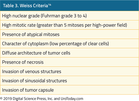
Investigation of suspected ACC should assess excesses of glucocorticoids, sex steroids, catecholamines, and mineralocorticoids. The Weiss pathologic criteria are used to distinguish benign and malignant adrenal lesions (Table 3).14 The presence of three or more of these criteria is highly associated with malignancy. 
table-3-approach-to-adrenal-masses.jpg

Treatment of Adrenal Lesions

For patients with small non-functional adrenal lesions with benign imaging findings, surveillance may be appropriate. However, surgery is the mainstay for patients with adrenal lesions. There are particular nuances on the basis of the underlying histology and functional status. In general, laparoscopic adrenalectomy is considered the gold standard as, in experienced hands, oncologic outcomes are equivalent with improved convalescence.

For patients with adrenocortical carcinoma, surgical resection is the standard of care. In these cases, wide margins are critical. Thus, for larger tumors with possible adjacent organ involvement, some authors advocate that these cases should be performed open in order to ensure negative margins given the potential need for adjacent organ resection. Unfortunately, recurrence is common following even aggressive resection. Radiotherapy can be used in an adjuvant setting for patients with positive margins and for treatment of bone or central nervous system metastases. Systemic therapy may be undertaken with mitotane, a synthetic derivative of DDT.

For patients with Cushing's disease, the management varies widely based on underlying etiology. The overall goals included correction of the cortisol excess, restoration of the underlying hormonal axis, and management of the sequelae. Approaches to this, depending on underlying etiology, include weaning of exogenous steroids, transsphenoidal resection of pituitary lesions, unilateral or bilateral adrenalectomy, resection of ectopic sources of ACTH, and medical therapy with blockers of steroidogenesis.

Treatment of primary aldosteronism seeks to control blood pressure and prevent sequelae of hormonal excess. This may be accomplished medically or surgically depending on the underlying cause and patient suitability for operation. Medical treatment may be undertaken with aldosterone receptor antagonists such as spironolactone or eplerenone.

Pheochromocytoma is primarily a surgical disease. However, extensive medical consultation and optimization is required to prevent significant intraoperative cardiovascular complications. Further, these patients are at risk of cardiomyopathy and, therefore, consultation with a cardiologist or anesthesiologist prior to surgery is advisable. Catecholamine blockade is required prior to surgery on pheochromocytoma. Classically, this has been achieved with the non-competitive alpha-blocker phenoxybenzamine. However, selective reversible alpha-blockers including doxazosin or terazosin are alternatives. Following alpha-blockade, beta-blockade may be undertaken due to the risk of reflex tachycardia or arrhythmia.13 An alternative to alpha- and beta-blockade which has been proposed utilized calcium channel blockade.15 Finally, catecholamine synthesis blockade through the use of alpha-methyltyrosine (metyrosine) may be added. In the perioperative period, repletion of the intravascular volume is critical. This may be achieved through liberal consumption of salt and liquid or intravenous resuscitation. Careful postoperative monitoring is key as these patients are at risk for hypotension and hypoglycemia. Additionally, as these lesions have a predilection for recurrence, ongoing monitoring is required.

Published Date: January 28th, 2019
References:
  1. Young WF, Jr. Clinical practice. The incidentally discovered adrenal mass. The New England journal of medicine 2007;356:601-10.
  2. Grumbach MM, Biller BM, Braunstein GD, et al. Management of the clinically inapparent adrenal mass ("incidentaloma"). Ann Intern Med 2003;138:424-9.
  3. Fassnacht M, Kroiss M, Allolio B. Update in adrenocortical carcinoma. The Journal of clinical endocrinology and metabolism 2013;98:4551-64.
  4. Lenert JT, Barnett CC, Jr., Kudelka AP, et al. Evaluation and surgical resection of adrenal masses in patients with a history of extra-adrenal malignancy. Surgery 2001;130:1060-7.
  5. Namimoto T, Yamashita Y, Mitsuzaki K, et al. Adrenal masses: quantification of fat content with double-echo chemical shift in-phase and opposed-phase FLASH MR images for differentiation of adrenal adenomas. Radiology 2001;218:642-6.
  6. Young WF, Jr., Klee GG. Primary aldosteronism. Diagnostic evaluation. Endocrinol Metab Clin North Am 1988;17:367-95.
  7. Bravo EL, Tagle R. Pheochromocytoma: state-of-the-art and future prospects. Endocr Rev 2003;24:539-53.
  8. Porterfield JR, Thompson GB, Young WF, Jr., et al. Surgery for Cushing's syndrome: an historical review and recent ten-year experience. World J Surg 2008;32:659-77.
  9. Newell-Price J, Bertagna X, Grossman AB, Nieman LK. Cushing's syndrome. Lancet 2006;367:1605-17.
  10. Lenders JW, Pacak K, Walther MM, et al. Biochemical diagnosis of pheochromocytoma: which test is best? JAMA : the Journal of the American Medical Association 2002;287:1427-34.
  11. Eisenhofer G, Goldstein DS, Walther MM, et al. Biochemical diagnosis of pheochromocytoma: how to distinguish true- from false-positive test results. The Journal of clinical endocrinology and metabolism 2003;88:2656-66.
  12. Motta-Ramirez GA, Remer EM, Herts BR, Gill IS, Hamrahian AH. Comparison of CT findings in symptomatic and incidentally discovered pheochromocytomas. AJR Am J Roentgenol 2005;185:684-8.
  13. Pacak K. Preoperative management of the pheochromocytoma patient. The Journal of Clinical Endocrinology and metabolism 2007;92:4069-79.
  14. Weiss LM. Comparative histologic study of 43 metastasizing and nonmetastasizing adrenocortical tumors. Am J Surg Pathol 1984;8:163-9.
  15. Ulchaker JC, Goldfarb DA, Bravo EL, Novick AC. Successful outcomes in pheochromocytoma surgery in the modern era. The Journal of Urology 1999;161:764-7.

Education Resources - Indwelling Catheters

This educational resource is a three-part series on the subject of catheter-associated urinary tract infections. In this webinar series, catheter-associated urinary tract infection as a part of the overall issue of hospital-acquired infection is examined. Why CAUTI is important and why this specific HAI has the attention of so many organizations and agencies is discussed. Finally, it examines the risk factors and pathogenesis for CAUTI, and what the current CDC definition of CAUTI is.

Systemic Therapy for Advanced Renal Cell Carcinoma

As highlighted in prior articles on the Etiology and Epidemiology of Kidney Cancer, cancers of the kidney and renal pelvis comprise the 6th most common newly diagnosed tumors in men and 10th most common in women1 and account for an estimated 65,340 people new diagnoses and 14,970 cancer-related deaths in 2018 in the United States. Despite a previously mentioned stage migration due to an increase in incidental detection, a large proportion (up to 35%) of patients present with advanced disease, including metastases.2 Historically, metastatic RCC has been early uniformly fatal, with 10-year survival rates less than 5%.3

As emphasized in the article on Malignant Renal Tumors, clear cell renal cell carcinoma (ccRCC) is the most common histologic subtype of renal cell carcinoma (RCC). Likely due to its much higher prevalence, the vast majority of systemic therapies for RCC have been investigated among patients with ccRCC. Historically, treatment for metastatic RCC (mRCC) had been limited to cytokine therapies (interleukin-2 and interferon-alfa). However, the development of tyrosine kinase inhibitors (TKIs), which target vascular endothelial growth factors (VEGF), and mammalian target of rapamycin (mTOR) inhibitors have replaced cytokine-based therapies as the standard of care. More recently, immunotherapy-based approaches using checkpoint inhibitors have demonstrated significant benefits and have joined the repertoire of available agents for patients with metastatic RCC.

Cytokine Therapies for Advanced ccRCC

The host immune system has long been implicated with RCC tumor biology. As a result, modulators of the immune system were among the first therapeutic targets for advanced ccRCC.

Interferon-α was one of the first cytokines assessed for the treatment of metastatic ccRCC. Interferons have a range of biologic functions, including immunomodulation. Early data demonstrated response rates in the range of 10 to 15% for patients treated with interferon-α.4 Compared with other available systemic therapies available at the time, interferon therapy conferred a survival benefit.5

An alternative form of immunologic modulation was examined using interleukin-2. While response rates were similar to interferon-based therapies (~15 to 20%)6, interleukin-2 was distinct in that durable complete responses were observed in approximately 7 to 9% of patients.7 On the basis of these data, high-dose IL-2 was approved by the U.S. Food and Drug Administration (FDA) in 1992. However, IL-2 is associated with considerable toxicity which has limited its widespread utilization. Most worrisome is vascular leak syndrome which leads to intravascular depletion, hypovolemia, respiratory compromise and multi-organ failure. Alternatives to the high-dose intravenous bolus administration were explored but lead to worse oncologic outcomes. Thus, high-dose IL-2 is the only recommended approach for patients undergoing cytokine therapy.

Subsequently, combinations of interferon and interleukin therapies were explored. These demonstrated some improvement in response rate but no difference in overall survival.8 Combination therapy resulted in significantly increased toxicity compared to monotherapy with either agent.

With the introduction of VEGF and mTOR targeting agents, interferon is no longer utilized as first-line therapy. However, IL-2 remains an available, though not widely utilized, option on the basis of its ability to induce durable complete responses which these new agents lack. 

Inhibitors of the VEGF Pathway for Advanced ccRCC

Based on work into the molecular biology underlying ccRCC led to “rational targeted therapeutics” including targeting of the VEGF pathway.

The first inhibitor of the VEGF pathway used in clinical trials was bevacizumab, a humanized monoclonal antibody against VEGF-A. While this approach was first explored in patients who had progressed on cytokine-based therapies, it was soon evaluated head-to-head against interferon in previously untreated patients.9,10 The addition of bevacizumab to interferon resulted in significant improvements in response rate and progression-free survival. Today, bevacizumab is uncommonly used as monotherapy in untreated patients but is considered as second-line therapy in patients who have failed prior therapy with tyrosine kinase inhibitors.

Tyrosine-kinase inhibitors also target the VEGF pathway, through inhibition of a combination of VEGFR-2, PDGFR-β, raf-1 c-Kit, and Flt3 (sunitinib and sorafenib). In 2006, sorafenib was shown to have biologic activity in ccRCC. Subsequent studies demonstrated improvements in progression-free survival compared with placebo in patients who have previously failed cytokine therapy and improvements in tumor regression compared to interferon in previously untreated patients. Despite FDA approval, sorafenib is rarely used as first-line therapy today. More widely used is sunitinib. As with agents discussed, sunitinib was first evaluated among patients who had previously received cytokine treatment. Subsequently, it was compared to interferon-α in a large phase III randomized trial.11 While the initial analysis demonstrated significant improvement in progression-free survival and overall response rate, subsequent follow-up has demonstrated a strong trend towards improved overall survival. On account of these data, sunitinib is widely used as first-line treatment of RCC. 

Tyrosine-kinase inhibitors exhibit a class-based toxicity profile including gastrointestinal events, dermatologic complications including hand-foot desquamation, hypertension, and general malaise. However, quality of life appears to be better with these agents than with interferon.11 

Subsequently, a number of more targeted tyrosine kinase inhibitors have been developed with the goal to decrease the toxicity of this treatment strategy. Such agents include pazopanib, axitinib, and tivozanib. Comparative data of pazopanib and sunitinib have demonstrated non-inferior oncologic outcomes with decreased toxicity among patients receiving pazopanib.12 Axitinib was evaluated as second-line therapy compared to sorafenib among patients who had previously received sunitinib, bevacizumab, temsirolimus, or cytokine therapy. Axitinib was associated with improved progression-free survival; on the basis of these data, this agent was approved for second-line therapy of metastatic RCC.13 Finally, tivozanib has been compared to sorafenib among patients who had not previously received VEGF or mTOR-targeting therapies. While this study demonstrated tivozanib’s activity, it was not FDA approved and is therefore not used.

Most recently, a multikinase inhibitor, cabozantinib, has been approved for the first-line treatment of mRCC. In the phase II CABOSUN trial, cabozantinib demonstrated improved progression-free survival compared to sunitinib.14 However, these results have proven controversial, with a number of concerns raised including a potential exaggerated effect due to the poor efficacy of sunitinib compared to what would be expected based on previous reports.15

Despite the efficacy of VEGF targeted therapies, resistance to VEGF-inhibition almost inevitably results. Therefore, research into the development of these resistance mechanisms and ways to target these pathways has been undertaken. Agents including nintedanib and dovitinib have been explored though these are not yet in routine practice.

Inhibitors of mTOR for Advanced ccRCC

Mammalian target of rapamycin (mTOR) plays a key role in regulating HIF-α, thus modulating the pathway between abnormalities in VHF and proliferation. Two analogous of sirolimus have demonstrated efficacy in treating advanced RCC, temsirolimus and everolimus.

A three-arm trial comparing temsirolimus, interferon, and the combination was undertaken among patients with pre-defined poor risk disease who had not previously received systemic therapy for RCC.16 This demonstrated improvements in progression-free survival and overall survival for patients receiving temsirolimus. Notably, the combination arm did not offer a benefit compared to interferon alone. Unlike temsirolimus which must be administered intravenously, everolimus is an oral agent. Among patients progressing on sunitinib and/or sorafenib, everolimus demonstrated significantly improved progression-free survival compared to placebo.17

Checkpoint Inhibitors for Advanced ccRCC

The immunologic basis for treatment of advanced RCC has been well established, including the aforementioned cytokine therapies. Recently, immune checkpoint inhibitors have been examined in the treatment of advanced RCC. Two particular regimes warrant focus – nivolumab and ipilimumab and atezolizumab.

First presented at ESMO in the fall of 2017 and subsequently published in the spring of 2018, CheckMate 214 demonstrated an overall survival (OS) benefit for first-line nivolumab plus ipilimumab vs. sunitinib.18 More details regarding this study may be found in the UroToday coverage of ESMO 2017. In short, among the subgroup of patients with intermediate or poor-risk RCC, treatment with nivolumab plus ipilimumab resulted in significantly improved overall response rate, comparable progression-free survival, and significantly improved overall survival.

Similarly, first presented at GU ASCO in the spring of 2018 and subsequently published, IMmotion151 reported a progression-free survival (PFS) benefit for first-line atezolizumab + bevacizumab vs. sunitinib.19 This regime was active with a significant benefit in progression-free survival among the whole cohort of patients, as well as a subset of PD-L1+ patients. More details regarding this study may be found in the UroToday coverage of GU ASCO 2018.

These trials are notable in that they demonstrated improved outcomes in first-line treatment, compared to the current standard of care, sunitinib.

Other Agents for Advanced ccRCC

Numerous chemotherapeutic agents have been explored in ccRCC. These include 5-FU, gemcitabine, vinblastine, bleomysin, and platinums. Meta-analyses of these data demonstrate poor response20 and thus cytotoxic chemotherapy is not indicated in the treatment of advanced RCC. Similarly, hormonal therapies including medroxyprogesterone have been explored but have no role in modern management of advanced RCC.

Treatment of Advanced non-ccRCC

There is a relative dearth of data for treatment of advanced non-clear cell RCC. Therefore, patients with these tumors may receive agents on the basis of their activity in ccRCC. However, VEGF-receptor inhibitors have been shown to have relatively low activity in patients with papillary RCC.21 Responses were somewhat better in patients with chromophobe RCC. Temsirolimus and everolimus appear to have some activity in patients with non-clear cell histology. Similarly, nivolumab monotherapy appears to have biologic activity in patients with non-ccRCC.22

Integration of treatment options for patients with advanced RCC

With so many active agents available for the treatment of advanced RCC, it may be difficult to ascertain which treatment to offer patients who present in clinic. There are a number of ways to approach this issue – first, one may take a quantitative approach, utilizing the available comparative data in a network meta-analysis; second, one may rely upon eminence, as in expert-informed guidelines; finally, one may rely on individual clinical experience. In this setting, all three options are available.

First, assessing this in a quantitative fashion, we performed a network meta-analysis of agents for the treatment of advanced RCC.23 While there are limitations to this approach including the reliance on the assumption of transitivity between studies, interesting conclusions may be drawn. First, assessing progression-free survival, we found that it was highly likely (91% chance) that cabozantinib provided the greatest benefit. However, when assessing overall survival, nivolumab plus ipilimumab had the highest likelihood of being the preferred treatment choice. Finally, when assessing adverse events, it was highly likely that nivolumab plus ipilimumab had the most favorable toxicity profile.

Second, considering a panel of expert opinion, the European Association of Urology updated their guidelines on the treatment of renal cell carcinoma recently. Their recommendations are highlighted in the following figure, taken from the EAU guidelines:
diagram-systemic-therapy@2x.jpg

Finally, we may rely on the guidance of individual clinical experience. Anil Kapoor, MD who has extensive experience in the treatment of both localized and advanced RCC, offered his treatment approach recently to UroToday.

Published Date: January 21st, 2019
 
References:
  1. Siegel RL, Miller KD, Jemal A. Cancer statistics, 2018. CA: a cancer journal for clinicians. 2018;68(1):7-30.
  2. Motzer RJ, Mazumdar M, Bacik J, Berg W, Amsterdam A, Ferrara J. Survival and prognostic stratification of 670 patients with advanced renal cell carcinoma. Journal of Clinical Oncology. 1999;17:2530-2540.
  3. Negrier S, Escudier B, Gomez F, et al. Prognostic factors of survival and rapid progression in 782 patients with metastatic renal carcinomas treated by cytokines: a report from the Groupe Francais d'Immunotherapie. Annals of oncology : official journal of the European Society for Medical Oncology / ESMO. 2002;13(9):1460-1468.
  4. Motzer RJ, Bacik J, Murphy BA, Russo P, Mazumdar M. Interferon-alfa as a comparative treatment for clinical trials of new therapies against advanced renal cell carcinoma. Journal of clinical oncology : official journal of the American Society of Clinical Oncology. 2002;20(1):289-296.
  5.  Coppin C, Porzsolt F, Awa A, Kumpf J, Coldman A, Wilt T. Immunotherapy for advanced renal cell cancer. Cochrane Database Syst Rev. 2005(1):CD001425.
  6. Dutcher JP, Atkins M, Fisher R, et al. Interleukin-2-based therapy for metastatic renal cell cancer: the Cytokine Working Group experience, 1989-1997. Cancer J Sci Am. 1997;3 Suppl 1:S73-78.
  7. Rosenberg SA, Yang JC, White DE, Steinberg SM. Durability of complete responses in patients with metastatic cancer treated with high-dose interleukin-2: identification of the antigens mediating response. Ann Surg. 1998;228(3):307-319.
  8. Negrier S, Escudier B, Lasset C, et al. Recombinant human interleukin-2, recombinant human interferon alfa-2a, or both in metastatic renal-cell carcinoma. Groupe Francais d'Immunotherapie. The New England journal of medicine. 1998;338(18):1272-1278.
  9. Rini BI, Halabi S, Rosenberg JE, et al. Bevacizumab plus interferon alfa compared with interferon alfa monotherapy in patients with metastatic renal cell carcinoma: CALGB 90206. Journal of clinical oncology: official journal of the American Society of Clinical Oncology. 2008;26(33):5422-5428.
  10. Escudier B, Pluzanska A, Koralewski P, et al. Bevacizumab plus interferon alfa-2a for treatment of metastatic renal cell carcinoma: a randomised, double-blind phase III trial. Lancet. 2007;370(9605):2103-2111.
  11. Motzer RJ, Hutson TE, Tomczak P, et al. Sunitinib versus interferon alfa in metastatic renal-cell carcinoma. The New England journal of medicine. 2007;356(2):115-124.
  12. Motzer RJ, Hutson TE, Cella D, et al. Pazopanib versus sunitinib in metastatic renal-cell carcinoma. The New England journal of medicine. 2013;369(8):722-731
  13. Rini BI, Escudier B, Tomczak P, et al. Comparative effectiveness of axitinib versus sorafenib in advanced renal cell carcinoma (AXIS): a randomised phase 3 trial. Lancet. 2011;378(9807):1931-1939.
  14. Choueiri TK, Halabi S, Sanford BL, et al. Cabozantinib Versus Sunitinib As Initial Targeted Therapy for Patients With Metastatic Renal Cell Carcinoma of Poor or Intermediate Risk: The Alliance A031203 CABOSUN Trial. Journal of clinical oncology: official journal of the American Society of Clinical Oncology. 2017;35(6):591-597
  15. Buti S, Bersanelli M. Is Cabozantinib Really Better Than Sunitinib As First-Line Treatment of Metastatic Renal Cell Carcinoma? Journal of clinical oncology: official journal of the American Society of Clinical Oncology. 2017;35(16):1858-1859.
  16.  Hudes G, Carducci M, Tomczak P, et al. Temsirolimus, interferon alfa, or both for advanced renal-cell carcinoma. The New England journal of medicine. 2007;356(22):2271-2281.
  17. Motzer RJ, Escudier B, Oudard S, et al. Efficacy of everolimus in advanced renal cell carcinoma: a double-blind, randomised, placebo-controlled phase III trial. Lancet. 2008;372(9637):449-456.
  18.  Escudier B, Tannir NM, McDermott D, et al. LBA5 - CheckMate 214: Efficacy and safety of nivolumab 1 ipilimumab (N1I) v sunitinib (S) for treatment-naive advanced or metastatic renal cell carcinoma (mRCC), including IMDC risk and PD-L1 expression subgroups. Annals of Oncology. 2017;28(Supplement 5):621-622.
  19.  Motzer R, Powles T, Atkins M, et al. IMmotion151: A Randomized Phase III Study of Atezolizumab Plus Bevacizumab vs Sunitinib in Untreated Metastatic Renal Cell Carcinoma. Journal of Clinical Oncology. 2018;36(Suppl 6S).
  20.  Yagoda A, Abi-Rached B, Petrylak D. Chemotherapy for advanced renal-cell carcinoma: 1983-1993. Semin Oncol. 1995;22(1):42-60.
  21. Choueiri TK, Plantade A, Elson P, et al. Efficacy of sunitinib and sorafenib in metastatic papillary and chromophobe renal cell carcinoma. Journal of clinical oncology : official journal of the American Society of Clinical Oncology. 2008;26(1):127-131
  22. Koshkin VS, Barata PC, Zhang T, et al. Clinical activity of nivolumab in patients with non-clear cell renal cell carcinoma. J Immunother Cancer. 2018;6(1):9.
  23. Wallis CJD, Klaassen Z, Bhindi B, et al. First-line Systemic Therapy for Metastatic Renal Cell Carcinoma: A Systematic Review and Network Meta-analysis. European urology. 2018;74(3):309-321.

Clinical Practice Inservice Tools - Indwelling Catheters

The following tools, outlining best practices in the handling of Foley catheters and urine drainage bag positioning, are available for download to support staff inservicing and other clinical practice educational initiatives:

Treatment of Metastatic Non-Clear Cell RCC

Background 

Kidney cancer is the 12th most common cancer in the world, with over 300,000 new cases annually, of which 65,340 new cases will be diagnosed in the United States in 2018.1 The incidence of renal cell carcinoma (RCC) varies substantially based on the country – rates of RCC are higher in Europe and North America and much lower in Asia and South America.2

Most kidney cancers (>90%) are renal cell carcinomas, and of renal cell carcinomas, the majority of cases (80%) will be the clear cell subtype.3 Of the remaining 20% of cases, the two major histological subtypes are papillary (10-14%) and chromophobe (5%)3,4, but also include collecting duct, translocation carcinoma, medullary carcinoma, and unclassified RCC. These histological subtypes are distinct from clear cell carcinoma and independently predict for survival.3 For example, after controlling for TNM status, age, gender, and tumor size, patients with early stage clear cell RCC are more than twice as likely to die of RCC than patients with papillary or chromophobe RCC.3 Some of these subtypes also have unique risk factors. For example, renal medullary carcinomas are an aggressive non-clear cell carcinoma that are almost exclusively associated with patients with sickle cell trait.5

diagram-1-treatment-non-clear-cell-RCC@2x.jpg

Non-clear cell RCCs (ncRCC) also have unique mutational landscapes.6 For example, MET mutations can be found in 15-30% of papillary RCCs.6,7 Papillary RCCs also have a higher mutation rate compared with chromophobe RCCs and renal oncocytomas. Durinck et al evaluated 167 primary human tumors including papillary, chromophobe and translocation subtypes and were able to use a five gene set to help molecularly distinguish between chromophobe carcinoma, renal oncocytoma and papillary carcinoma.6

Treatment

Given the rarity of non-clear cell RCC (ncRCC), there is a paucity of large randomized phase III trials to help guide the optimal therapy for ncRCC. Some trials for ncRCC have had to stop due to slow accrual. Given this lack of data, patients are encouraged to participate in clinical trials when they are available and appropriate. Below is a summary of the most common systemic therapies in use for ncRCC.
table-1-treatment-non-clear-cell-RCC@2x.jpg

Sunitinib

Single agent sunitinib has been evaluated as part of an expanded access trial as well as several small phase II trials (Table 1). In a single arm phase II clinical trial, 23 patients were given sunitinib 50 mg in cycles of four weeks on followed by 2 weeks off. The trial was stopped early due to slow accrual and the median progression free survival (PFS) was 5.5 months.8  In a another phase II trial evaluating both sunitinib and sorafenib, 19 patients with ncRCC were given sunitinib – median PFS was 11.9 months in the papillary arm and 8.9 months in the chromophobe arm.9 The largest of these studies was reported by Gore et al, which included 588 ncRCC patients who received sunitinib as part of the expanded access study encompassing 4564 RCC patients who received sunitinib.10 In their study, 11% of patients had an objective response and median PFS was 7.8 months. Some trials have stratified outcomes by histological subtypes such as chromophobe or papillary, and one trial reported results broken down by type 1 papillary RCC vs type 2 papillary RCC11. Overall, most studies demonstrated that sunitinib led to a median PFS of about 6-7 months for ncRCC.
table-2-treatment-non-clear-cell-RCC@2x.jpg

Everolimus and Temsirolimus

mTOR inhibitors Everolimus and Temsirolimus have been evaluated by a few phase II trials for ncRCC (Table 2). Everolimus was evaluated in a subgroup of REACT (RAD001 Expanded Access Clinical Trial in RCC) and in RAPTOR (RAD001 in Advanced Papillary Tumor Program in Europe).15,16 In REACT, 1.3% of patients with ncRCC had a partial response and 49.3% had stable disease.15 In RAPTOR, the median progression free survival (mPFS) was 4.1 months and median OS was 21.4 months. For patients with chromophobe RCC, mTOR directed therapy may be especially effective – case reports show partial responses and stable disease to both Everolimus or Temsirolimus.17,18 This may be due to the hypothesis that a high number of chromophobe RCC’s have PI3K-mTOR pathway activation as well as frequent TSC1/TSC2 mutations which may sensitize these tumors to mTOR inhibition.19 

Sunitinib vs Everolimus

Sunitinib and everolimus have also been examined head to head. The largest trial was ASPEN (Everolimus versus sunitinib for patients with metastatic non-clear-cell renal cell carcinoma), a multicenter open-label, randomized phase II trial which randomized 108 patients to receive either sunitinib or everolimus.22 The primary endpoint of this study was progression free survival. The majority of patients had papillary histology (65%). Median overall survival was 13.2 months in the everolimus group and 31.5 months in the sunitinib group. However, overall survival was not statistically different between the two treatment groups (HR 1.12, 95%CI 0.7-2.1, p=0.6). A second study, ESPN (Everolimus Versus Sunitinib Prospective Evaluation in Metastatic Non–Clear Cell Renal Cell Carcinoma) came to a similar conclusion and found that the median overall survival was 16.2 months with sunitinib and 14.9 months with everolimus (p=0.18).23

While overall survival between everolimus and sunitinib were not statistically different for the unselected cohorts, ASPEN did find differences in objective responses between the different subtypes, suggesting that each subtype of ncRCC may respond differently to therapies. In ASPEN, 24% (8/33) of papillary RCCs achieved a partial or complete radiographic response on sunitinib compared with 5% of patients on everolimus (2/37). Interestingly, clinical outcomes after receipt of either sunitinib or everolimus also varied based on risk stratification. Patients with good or intermediate risk had improved median progression free survival (mPFS) with first line sunitinib than everolimus. However, patients with poor risk had improved PFS with everolimus over sunitinib. This is concurrent with the ARCC trial, which also demonstrated improved overall survival with an mTOR inhibitor (temsirolimus) in poor risk patients with ncRCC.24

A meta-analysis of five studies (ESPN, ASPEN, RECORD3, ARCC, and SWOG1107) found a nonstatistical trend favoring sunitinib over everolimus for ncRCC but does note that there is considerable patient heterogeneity in these small studies and there was no statistical difference in PFS between these two therapies.25 In the absence of clinical trial options, sunitinib is a reasonable first line choice for treatment naïve patients with ncRCC, especially for those with papillary RCC or MSKCC good risk RCC.

Special Populations

It is well recognized that ncRCC is a heterogenous mix of patients which respond differently to therapies. Thus, there have been a few biomarker or histology driven trials looking at specific subsets of ncRCC. For example, for patients with hereditary leiomyomatosis and papillary RCC, a phase II study of 41 patients found that patients treated bevacizumab plus erlotinib had a median PFS of 24.2 months, compared to 7.4 months for patients with sporadic papillary RCC.26 Patients with papillary RCC frequently have MET mutations and a variety of MET inhibitors including crizotinib, savolitinib, and cabozantinib are being evaluated in clinical trials.27-29 For example, in a study of 41 patients with type 1 papillary RCC, of the 4 patients with MET+ tumors, 2 had achieved a partial response and one had stable disease with crizotinib.28 In a study with Savolitinib, another selective MET inhibitor, patients with MET “driven” tumors had a median PFS of 6.2 months, compared with 1.7 months for MET independent tumors.29

table-3-treatment-non-clear-cell-RCC@2x.jpg

Future Direction

A number of active clinical trials are in progress, investigating various MET inhibitors as well as checkpoint inhibitors for ncRCC (Table 3). Preliminary data suggest that PD-1 or PD-L1 blockade may have some activity in this population.30,31 Given the dearth of data and rarity of ncRCC, it is important to consider these patients for clinical trials, whenever possible.

Published Date: November 29th, 2018

Written by: Jason Zhu, MD
References:

1. Motzer R, Jonasch E, Agarwal N. Kidney Cancer: NCCN Evidence Blocks, Version 2.2018, NCCN Clinical Practice Guidelines in Oncology. 2017.

2. Chow W-H, Dong LM, Devesa SS. Epidemiology and risk factors for kidney cancer. Nature Reviews Urology 2010;7:245.

3. Leibovich BC, Lohse CM, Crispen PL, et al. Histological Subtype is an Independent Predictor of Outcome for Patients With Renal Cell Carcinoma. The Journal of Urology 2010;183:1309-16.

4. Moch H, Gasser T, Amin MB, Torhorst J, Sauter G, Mihatsch MJ. Prognostic utility of the recently recommended histologic classification and revised TNM staging system of renal cell carcinoma. Cancer 2000;89:604-14.

5. Shetty A, Matrana MR. Renal Medullary Carcinoma: A Case Report and Brief Review of the Literature. The Ochsner Journal 2014;14:270-5.

6. Durinck S, Stawiski EW, Pavía-Jiménez A, et al. Spectrum of diverse genomic alterations define non–clear cell renal carcinoma subtypes. Nature genetics 2015;47:13.

7. Carlo MI, Khan N, Chen Y, et al. The genomic landscape of metastatic non-clear cell renal cell carcinoma. American Society of Clinical Oncology; 2017.

8. Molina AM, Feldman DR, Ginsberg MS, et al. Phase II trial of sunitinib in patients with metastatic non-clear cell renal cell carcinoma. Investigational New Drugs 2012;30:335-40.

9. Choueiri TK, Plantade A, Elson P, et al. Efficacy of Sunitinib and Sorafenib in Metastatic Papillary and Chromophobe Renal Cell Carcinoma. Journal of Clinical Oncology 2008;26:127-31.

10. Gore ME, Szczylik C, Porta C, et al. Safety and efficacy of sunitinib for metastatic renal-cell carcinoma: an expanded-access trial. The Lancet Oncology 2009;10:757-63.

11. Ravaud A, Oudard S, De Fromont M, et al. First-line treatment with sunitinib for type 1 and type 2 locally advanced or metastatic papillary renal cell carcinoma: a phase II study (SUPAP) by the French Genitourinary Group (GETUG)†. Annals of Oncology 2015;26:1123-8.

12. Tannir NM, Plimack E, Ng C, et al. A phase 2 trial of sunitinib in patients with advanced non–clear cell renal cell carcinoma. European urology 2012;62:1013-9.

13. Lee JL, Ahn JH, Lim HY, et al. Multicenter phase II study of sunitinib in patients with non-clear cell renal cell carcinoma. Annals of Oncology 2012;23:2108-14.

14. Shi H-Z, Tian J, Li C-L. Safety and efficacy of sunitinib for advanced non-clear cell renal cell carcinoma. Asia-Pacific Journal of Clinical Oncology 2015;11:328-33.

15. Blank CU, Bono P, Larkin JMG, et al. Safety and efficacy of everolimus in patients with non-clear cell renal cell carcinoma refractory to VEGF-targeted therapy: Subgroup analysis of REACT. Journal of Clinical Oncology 2012;30:402-.

16. Escudier B, Molinie V, Bracarda S, et al. Open-label phase 2 trial of first-line everolimus monotherapy in patients with papillary metastatic renal cell carcinoma: RAPTOR final analysis. European Journal of Cancer 2016;69:226-35.

17. Larkin JMG, Fisher RA, Pickering LM, et al. Chromophobe Renal Cell Carcinoma With Prolonged Response to Sequential Sunitinib and Everolimus. Journal of Clinical Oncology 2011;29:e241-e2.

18. Shuch B, Vourganti S, Friend JC, Zehngebot LM, Linehan WM, Srinivasan R. Targeting the mTOR pathway in chromophobe kidney cancer. Journal of Cancer 2012;3:152.

19. Maroto P, Anguera G, Roldan-Romero JM, et al. Biallelic TSC2 Mutations in a Patient With Chromophobe Renal Cell Carcinoma Showing Extraordinary Response to Temsirolimus. Journal of the National Comprehensive Cancer Network 2018;16:352-8.

20. Dutcher JP, Atkins M, Fisher R, et al. Interleukin-2-based therapy for metastatic renal cell cancer: the Cytokine Working Group experience, 1989-1997. The cancer journal from Scientific American 1997;3:S73-8.

21. Koh Y, Lim HY, Ahn JH, et al. Phase II trial of everolimus for the treatment of nonclear-cell renal cell carcinoma. Annals of Oncology 2013;24:1026-31.

22. Armstrong AJ, Halabi S, Eisen T, et al. Everolimus versus sunitinib for patients with metastatic non-clear cell renal cell carcinoma (ASPEN): a multicentre, open-label, randomised phase 2 trial. The Lancet Oncology 2016;17:378-88.

23. Tannir NM, Jonasch E, Albiges L, et al. Everolimus versus sunitinib prospective evaluation in metastatic non–clear cell renal cell carcinoma (ESPN): a randomized multicenter phase 2 trial. European urology 2016;69:866-74.

24. Hudes G, Carducci M, Tomczak P, et al. Temsirolimus, Interferon Alfa, or Both for Advanced Renal-Cell Carcinoma. New England Journal of Medicine 2007;356:2271-81.

25. Fernández-Pello S, Hofmann F, Tahbaz R, et al. A systematic review and meta-analysis comparing the effectiveness and adverse effects of different systemic treatments for non-clear cell renal cell carcinoma. European urology 2017;71:426-36.

26. Srinivasan R, Su D, Stamatakis L, et al. 5 Mechanism based targeted therapy for hereditary leiomyomatosis and renal cell cancer (HLRCC) and sporadic papillary renal cell carcinoma: interim results from a phase 2 study of bevacizumab and erlotinib. European Journal of Cancer 2014;50:8.

27. Martinez Chanza N, Bossé D, Bilen MA, et al. Cabozantinib (Cabo) in advanced non-clear cell renal cell carcinoma (nccRCC): A retrospective multicenter analysis. American Society of Clinical Oncology; 2018.

28. Schoffski P, Wozniak A, Escudier B, et al. Effect of crizotinib on disease control in patient with advanced papillary renal cell carcinoma type 1 with MET mutations or amplification: Final results of EORTC 90101 CREATE. American Society of Clinical Oncology; 2018.

29. Choueiri TK, Plimack ER, Arkenau H-T, et al. A single-arm biomarker-based phase II trial of savolitinib in patients with advanced papillary renal cell cancer (PRCC). American Society of Clinical Oncology; 2017.

30. Moreira RB, McKay RR, Xie W, et al. Clinical activity of PD1/PDL1 inhibitors in metastatic non-clear cell renal cell carcinoma (nccRCC). American Society of Clinical Oncology; 2017.

31. Chahoud J, Campbell MT, Gao J, et al. Nivolumab (nivo) for patients (pts) with metastatic non-clear cell renal cell carcinoma (nccRCC): A single-institution experience. American Society of Clinical Oncology; 2018.

Education Tools - Indwelling Catheters

The following tools, outlining best practices in the handling of Foley catheters and urine drainage bag positioning, are available for download:

care and use of foley330c

download button
 

drainage bag330c

download button


indications iuc330d

download button
 

summary330

download button


fastfacts330b

download button


Published Date: January 2013

Epidemiology and Etiology of Kidney Cancer

Kidney cancer is a broad, encompassing term that borders on colloquial. While most physicians are referring to renal cell carcinoma when they say “kidney cancer”, a number of other benign and malignant lesions may similarly manifest as a renal mass. Considering only the malignant causes, kidney cancers may include renal cell carcinoma, urothelium-based cancers (including urothelial carcinoma, squamous cell carcinoma, and adenocarcinoma), sarcomas, Wilms tumor, primitive neuroectodermal tumors, carcinoid tumors, hematologic cancers (including lymphoma and leukemia), and secondary cancers (i.e. metastases from other solid organ cancers).

Epidemiology

In the United States, cancers of the kidney and renal pelvis comprise the 6th most common newly diagnosed tumors in men and 10th most common in women.1 In 2018, an estimated 65,340 people will be newly diagnosed with cancers of the kidney and renal pelvis in the United States. In men, this comprises 42,680 estimated new cases in 2018 representing 5% of all newly diagnosed cancers. In women, 22,660 new cases are anticipated in 2018 representing 3% of all newly diagnosed cancers. Additionally, 14,970 people are expected to die of kidney and renal pelvis cancers in 2018 in the United States, with this being the 10th most common cause of oncologic death among men.

In Europe, results are similar. In 2018, the incidence of kidney cancer is estimated at 136,500 new cases representing 3.5% of all new cancer diagnoses.2 This corresponds to an estimated age standardized rate (ASR) of 13.3 cases per 100,000 population. As in the United States, the incidence of kidney and renal pelvis cancers is higher among men (incidence 84,9000, 4.1% of all cancers, ASR 18.6 per 100,000) than women (incidence 51,600, 2.8% of all cancers, ASR 9.0 per 100,000). Correspondingly, 54,700 people were estimated to die of kidney and renal pelvis cancers in Europe in 2018, accounting for 2.8% of all oncologic deaths. The age standardized mortality rate was 4.7 deaths per 100,000 population. Again, death from kidney and renal pelvis cancer was more common among men (mortality 35,100, 3.3% of oncologic deaths, ASR 7.1 per 100,000) than among women (mortality 19,600, 2.3% of oncologic deaths, ASR 2.7 per 100,000). Interestingly, within Europe, there is considerable variation in the incidence and mortality of kidney and renal pelvis cancer between countries.2

While the aforementioned data have already demonstrated that gender is strongly associated with the risk of both diagnosis of and death from kidney and renal pelvis cancers, age also importantly moderates this risk. Among patients in the United States, the probability of developing kidney and renal pelvis cancer rises nearly ten fold from age <50 to age >70 years.1

table 1 epidemiology kidney cancer2x
Thus, kidney cancer is predominantly a disease of older adults, with the typical presentation being between 50 and 70 years of age. However, over time, rates of diagnosis of kidney cancer have increased fastest among patients aged less than 40 years old.3

In the United States, kidney cancers are more common among African Americans, American Indians, and Alaska Native populations while rates are lower among Asian Americans.4 Worldwide, the highest rates are found in European nations while low rates are seen in African and Asian countries.4

The vast majority of patients have localized disease at the time of presentation. According to Siegel et al., 65% of all patients diagnosed with kidney and renal pelvis tumors between 2007 and 2013 had localized disease at the time of presentation while 16% had regional spread and 16% had evidence of distant, metastatic disease.1 This is in large part due to incidental diagnosis due to the increased use of ultrasonography and computed tomography in patients presenting with abdominal distress. In fact, 13 to 27% of abdominal imaging studies demonstrate incidental renal lesions unrelated to the reason for the study5 and approximately 80% of these masses are malignant.6 Dr. Welch and colleagues demonstrated elegantly that the use of computed tomography is strongly related to the likelihood of undergoing nephrectomy, likely due to detection of renal masses. Thus, with the increasing utilization of abdominal imaging, the incidence of kidney cancer has increased by approximately 3 to 4% per year since the 1970s.

Renal Cell Carcinoma

Renal cell carcinoma (RCC) is the most common kidney cancer. A number of histological subtypes have been recognized including conventional clear cell RCC (ccRCC), papillary RCC, chromophobe RCC, collecting duct carcinoma, renal medullary carcinoma, unclassified RCC, RCC associated with Xp11.2 translocations/TFE3 gene fusions, post-neuroblastoma RCC, and mucinous tubular and spindle cell carcinoma. Conventional ccRCC comprises approximately 70-80% of all RCCs while papillary RCC comprises 10-15%, chromophobe 3-5%, collecting duct carcinoma <1%, unclassified RCC 1-3%, and the remainder are very uncommon.

Histologically, most of these tumors are believed to arise from the cells of the proximal convoluted tubule given their ultrastructural similarities. Renal medullary carcinoma and collecting duct carcinoma, relatively uncommon and aggressive subtypes of RCC, are believed to arise more distally in the nephron.

Familial RCC Syndromes

While the vast majority of newly diagnosed RCCs are sporadic, hereditary RCCs account for approximately 4% of all RCCs. Due in large part to the work of Dr. Linehan and others, the understanding of the underlying molecular genetics of RCC have progressed significantly since the early 1990s. These insights have led to a better understanding of both familial and sporadic RCCs.

Von Hippel-Lindau disease is the most common cause of hereditary RCC. Due to defects in the VHL tumor suppressor gene (located at 3p25-26), this syndrome is characterized by multiple, bilateral clear cell RCCs, retinal angiomas, central nervous system hemangioblastomas, pheochromocytomas, renal and pancreatic cysts, inner ear tumors, and cystadenomas of the epididymis. RCC develops in approximately 50% of individuals with VHL disease. These tumors are characterized by an early age at the time of diagnosis, bilaterality, and multifocality. Due in large part to improved management of the CNS disorders in VHL disease, RCC is the most common cause of death in patients with VHL.

Hereditary papillary RCC (HPRCC) is, as one would expect from the name, associated with multiple, bilateral papillary RCCs. Due to an underlying constitutive activation of the c-Met proto-oncogene (located at 7q31), these tumors also present at a relatively early age. However, overall, these tumors appear in general to be less aggressive than corresponding sporadic malignancies.

In contrast, tumors arising in hereditary/familial leiomyomatosis and RCC (HLRCC), due to a defect in the fumarate hydratase (1q42-43) tumor suppressor gene, are typically unilateral, solitary, and aggressive. Histologically, these are typically type 2 papillary RCC, which has a more aggressive phenotype, or collecting duct carcinomas. Extra-renal manifestations include leiomyomas of the skin and uterus and uterine leiomyosarcomas which contribute to the name of this sydrome.

Birt-Hogg-Dube, due to defect in the tumor suppressor folliculin (17p11), is associated with multiple chromophobe RCCs, hybrid oncocytic tumors (with characteristics of both chromophobe RCC and oncocytoma), oncocytoma. Less commonly, patients with Birt-Hogg-Dube may develop clear cell RCC or papillary RCC. Non-renal manifestations include facial fibrofolliculomas, lung cysts, and the development of spontaneous pneumothorax.

Tuberous sclerosis, due to defects in TSC1 (located at 9q34) or TSC2 (16p13), may lead to clear cell RCC. More commonly, it is associated with multiple benign renal angiomyolipomas, renal cystic disease, cutaneous angiofibromas, and pulmonary lymphangiomyomatosis.

Succinate dehydrogenase RCC, due to defects in the SDHB (1p36.1-35) or SDHD (11q23) subunits of the succinate dyhydrogenase complex, may lead to a variety of RCC subtypes including chromophobe RCC, clear cell RCC, and type 2 papillary RCC. Extra-renal manifestations including benign and malignant paragangliomas and papillary thyroid carcinoma. In general, these tumors exhibit aggressive behaviour and wide surgical excision is recommended.

Finally, Cowden syndrome, due to defects in PTEN (10q23) may lead to papillary or other RCCs in addition to benign and malignant breast tumors and epithelial thyroid cancers.

Etiologic Risk Factors in Sporadic RCC

While numerous hereditary RCC syndromes exist, they account for only 4% of all RCCs. However, many sporadic RCCs share similar underlying genetic changes including VHL defects in ccRCC and c-Met activation in papillary RCC. A number of modifiable risk factors associated with RCC have been described.4

The foremost risk factor for the development of RCC is cigarette smoking. According to both the US Surgeon General and the International Agency for Research on Cancer, observational evidence is sufficient to conclude there is a causal relationship between tobacco smoking and RCC. A comprehensive meta-analysis of western populations demonstrated an overall relative risk for the development of RCC of 1.38 (95% confidence interval 1.27 to 1.50) for ever smokers compared to lifetime never smokers.7 Interestingly, this effect was larger for men (RR 1.54, 95% CI 1.42-1.68) than for women (RR 1.22, 95% CI 1.09-1.36). Additionally, there was a strong dose response relationship: compared to never smokers, men who smoked 1-9 cigarettes per day had a 1.6x risk, those who smoked 10-20 per days had a 1.83x risk, and those who smoked more than 21 per day had a 2.03x risk. A similar trend was seen among women. Notably, the risk of RCC declined with increasing durations of abstinence of smoking. Smoking appears to be preferentially associated with the development of clear cell and papillary RCC.8 In addition to being associated with increased RCC incidence, smoking is associated with more aggressive forms of RCC, manifest with higher pathological stage and an increased propensity for lymph node involvement and metastasis at presentation.9 As a result, smokers have worse cancer-specific and overall survival.9

Second, obesity is associated with an increased risk of RCC. While this risk was historically felt to be higher among women, a more recent review demonstrated no such effect modification according to sex.10 In a meta-analysis of 22 studies, Bergstrom et al. estimated that each unit increase of BMI was associated with a 7% increase in the relative risk of RCC diagnosis.

Third, hypertension has been associated with an increased risk of RCC diagnosis, with a hazard ratio of 1.70 (95%CI 1.30-2.22) in the VITAL study.11 Interestingly, in an American multiethnic cohort, this effect appeared to be larger among women (RR 1.58, 95% CI 1.09-2.28) than in men (RR 1.42, 95% CI 1.07-1.87).12 Again, as with obesity, there appears to be a dose-effect relationship between severity of hypertension and the risk of RCC diagnosis.13

Fourth, acquired cystic kidney disease (ACKD) appears to be associated with a nearly 50x increase risk of RCC diagnosis.14 ACKD occurs in patients with end-stage renal disease on dialysis. These changes are common among patients who have been on dialysis for at least 3 years.14 Interestingly, the risk of RCC appears to decrease following renal transplantation.

Finally, a number of other putative risk factors have been described. These lack the voracity of data that the aforementioned four have. Such risk factors include alcohol, analgesics, diabetes, and diet habits.4

Published Date: November 20th, 2018

References:

1. Siegel RL, Miller KD, Jemal A. Cancer statistics, 2018. CA: a cancer journal for clinicians 2018;68:7-30.

2. Ferlay J, Colombet M, Soerjomataram I, et al. Cancer incidence and mortality patterns in Europe: Estimates for 40 countries and 25 major cancers in 2018. European journal of cancer 2018.

3. Nepple KG, Yang L, Grubb RL, 3rd, Strope SA. Population based analysis of the increasing incidence of kidney cancer in the United States: evaluation of age specific trends from 1975 to 2006. The Journal of urology 2012;187:32-8.

4. Kabaria R, Klaassen Z, Terris MK. Renal cell carcinoma: links and risks. Int J Nephrol Renovasc Dis 2016;9:45-52.

5. Gill IS, Aron M, Gervais DA, Jewett MA. Clinical practice. Small renal mass. The New England journal of medicine 2010;362:624-34.

6. Frank I, Blute ML, Cheville JC, Lohse CM, Weaver AL, Zincke H. Solid renal tumors: an analysis of pathological features related to tumor size. The Journal of urology 2003;170:2217-20.

7. Hunt JD, van der Hel OL, McMillan GP, Boffetta P, Brennan P. Renal cell carcinoma in relation to cigarette smoking: meta-analysis of 24 studies. International journal of cancer Journal international du cancer 2005;114:101-8.

8. Patel NH, Attwood KM, Hanzly M, et al. Comparative Analysis of Smoking as a Risk Factor among Renal Cell Carcinoma Histological Subtypes. The Journal of urology 2015;194:640-6.

9. Kroeger N, Klatte T, Birkhauser FD, et al. Smoking negatively impacts renal cell carcinoma overall and cancer-specific survival. Cancer 2012;118:1795-802.

10. Bergstrom A, Hsieh CC, Lindblad P, Lu CM, Cook NR, Wolk A. Obesity and renal cell cancer--a quantitative review. British journal of cancer 2001;85:984-90.

11. Macleod LC, Hotaling JM, Wright JL, et al. Risk factors for renal cell carcinoma in the VITAL study. The Journal of urology 2013;190:1657-61.

12. Setiawan VW, Stram DO, Nomura AM, Kolonel LN, Henderson BE. Risk factors for renal cell cancer: the multiethnic cohort. American journal of epidemiology 2007;166:932-40

13. Vatten LJ, Trichopoulos D, Holmen J, Nilsen TI. Blood pressure and renal cancer risk: the HUNT Study in Norway. British journal of cancer 2007;97:112-4.

14. Brennan JF, Stilmant MM, Babayan RK, Siroky MB. Acquired renal cystic disease: implications for the urologist. Br J Urol 1991;67:342-8.