BERKELEY, CA (UroToday.com) - The use of focused bedside diagnostic ultrasound performed by non-radiologists, such as emergency physicians, is on the increase worldwide.[1] As ultrasound equipment has become portable and easier to use, various organizations have endorsed training and certification programs that bring a limited set of diagnostic skills to the physician making clinical decisions at the bedside.
The American College of Emergency Physicians (ACEP) offered its first course specifically dedicated to training emergency providers in the use of bedside ultrasound in 1990. In 1994, the Society of Academic Emergency Medicine (SAEM) released its model curriculum for physician training in emergency ultrasonography, which consisted of physics and instrumentation, cardiovascular, abdominal, and obstetric and gynecologic applications.[2] The scope and practice of clinical ultrasound by emergency physicians has been expanding gradually to include an increased number of indications and techniques (Table 1).
Table 1. Current emergency bedside ultrasound applications[3]
|
Core Applications |
Emerging/Adjunct Applications |
|
Trauma Intrauterine pregnancy AAA Cardiac Biliary Urinary Tract DVT Soft-tissue/musculoskeletal Thoracic Ocular Procedural guidance |
Advanced echo Transesophageal echo Bowel Adnexal pathology Testicular Transcranial Doppler Contrast studies |
Adapted from American College of Emergency Physicians Policy Statement: Emergency Ultrasound Guidelines, October 2008.
Focused bedside ultrasound examinations differ from traditional radiology studies in many ways, but in essence, their purpose is far more specific and limited. Ultrasound examinations performed in the emergency department are meant to answer one or two simple clinical questions that are critical for clinical decision making. The answers to these questions tend to be binary, i.e., is there a fetus in the uterus or not? Does this patient have hydronephrosis or not? Moreover, because of the limited nature of these ultrasound examinations, emergency physicians are taught to “rule in” important pathologies or findings, such as “positive free fluid in the peritoneum” rather than to rule them out. In other words, bedside ultrasound is used as a tool to expedite care when certain findings are present, not as a definitive modality to eliminate certain diagnoses. Patients in the emergency department, where bedside ultrasound is in everyday use by emergency providers, are also sometimes sent to the radiology department for complete or “formal” ultrasound examinations in the course of their care.
All residents training in emergency medicine in the United States receive training in emergency ultrasound. As part of the curriculum outlined by SAEM, residents receive 40 hours of didactic instruction and are expected to complete a total of 150 proctored examinations. In addition, there are currently 62 fellowship programs in the United States in emergency ultrasound that offer training for emergency physicians to obtain further expertise in the modality. These are typically one or two years in duration.
Renal colic is widely considered to be one of the primary indications for emergency department focused ultrasound. In fact, genitourinary ultrasound is often the first examination taught in bedside ultrasound courses. With the patient supine, a 3.5-5 MHz probe is placed in the right lower intercostal space, mid-axillary line. The probe is gently rocked to fan through the entire kidney while obtaining longitudinal and transverse views. The left kidney is imaged with the probe in the posterior axillary line. The degrees of hydronephrosis are seen in Figure 1.
Figure 1. Degrees of Hydronephrosis.[4]
Source: Emory University School of Medicine: http://www.em.emory.edu/ultrasound/ImageWeek/hydronephrosis.html
Non-enhanced helical computed tomography (CT) is commonly used to aid the diagnosis of nephrolithiasis, but it carries with it significant cost, incidental findings, and ionizing radiation. From 1996 to 2007, there was a 10-fold increase in the utilization of CT scan for patients with suspected kidney stone without an associated change in the proportion of diagnosis of kidney stone, significant alternate diagnoses, or admission to the hospital.[5] Ultrasound can be performed safely and quickly at the bedside with essentially no risks.
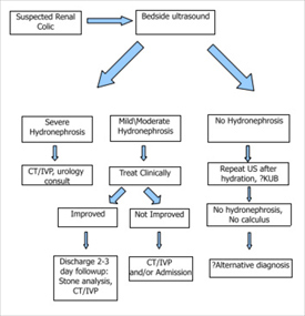
In the late 1990s, we proposed an algorithm for the management of renal colic in emergency department patients.[6] Noble and Brown and then Kartal and colleagues both attempted to validate a similar clinical algorithm. Kartal’s group was able to discharge over 50% of patients who presented with renal colic with no further imaging in the ED other than a bedside ultrasound performed by an emergency clinician. In patients presenting with acute flank pain in whom renal colic was suspected, those patients with hematuria and hydronephrosis on bedside ultrasound were discharged without further imaging in the emergency department. There were no serious adverse events at 2 months of follow-up in this group.[7]
Figure 2. An algorithm for the management of suspected renal colic in the emergency department.[8]
Peregrine JD, Noble VE. Bedside ultrasound and the assessment of renal colic. Emerg Med J. 2013;30(1):3-8
Our research could be of benefit here by incorporating the finding of hematuria (microscopic or macroscopic) into clinical decision making. In our cohort, no patient without hydronephrosis on bedside ultrasound and without hematuria had a stone larger than 5mm. This is a significant threshold, as stones greater than 5mm pass much less commonly without intervention. In someone for whom a stone is very likely, like a young male who previously had stones, but presents without hydronephrosis and no hematuria, the patient may indeed have a stone, but not one >5mm.
Although there is still no prospectively validated scoring system or algorithm widely in use, such a strategy could greatly improve the emergency management of patients with suspected renal colic by reducing unnecessary imaging, saving money to the health system, and reducing the radiation dose to patients who will not benefit from it.
Clinicians must keep in mind that other pathological processes present with flank pain. Bedside ultrasound in the emergency department is not used to identify life-threatening alternative diagnoses like bowel obstructions, appendicitis, diverticulitis, and ovarian torsion. These diagnoses cannot be missed. It is important to recognize the role of CT in diagnosing these pathologies. Astute clinicians will also use bedside ultrasound to evaluate for abdominal aortic aneurysm.
We wait with anticipation the results of the STONE trial, a prospective multi-centered study that has recently finished enrolling >2700 emergency department patients with suspected renal colic. As the data continues to accumulate, ultrasound continues to cement itself as the first line imaging modality for emergency department patients with suspected nephrolithiasis.
References:
- Cardenas E. Emergency medicine ultrasound policies and reimbursement guidelines. Emerg Med Clin North Am 2004; 22:829–838, 10-11.
- Mateer J, Plummer D, Heller M, Olson D, Jehle D, Overton D, Gussow L. Model curriculum for physician training in emergency ultrasonography. Ann Emerg Med. 1994 Jan;23(1):95-102.
- American College of Emergency Physicians Policy Statement: Emergency Ultrasound Guidelines, October 2008.
- Emory University School of Medicine Department of Emergency Medicine website: http://www.em.emory.edu/ultrasound/ImageWeek/hydronephrosis.html
- Westphalen A, Hsia RY, Maselli JH, et al. Radiological imaging of patients with suspected urinary tract stones: national trends, diagnoses, and predictors. Acad Emerg Med 2011;18:700–7.
- Swadron S, Mandavia DP. Renal Ultrasound. In: Ma OJ, Mateer JR, editors. Emergency Ultrasound. New York: McGraw Hill Professional; 2002. Pp.197-220
- Kartal M, Eray O, Erdogru T, et al. Prospective validation of a current algorithm including bedside US performed by emergency physicians for patients with acute flank pain suspected for renal colic. Emerg Med J 2006; 23:341–4.
- Peregrine JD, Noble VE. Bedside ultrasound and the assessment of renal colic. Emerg Med J. 2013;30(1):3-8
Written by:
Jeff Riddell* and Stuart Swadron** as part of Beyond the Abstract on UroToday.com. This initiative offers a method of publishing for the professional urology community. Authors are given an opportunity to expand on the circumstances, limitations etc... of their research by referencing the published abstract.
* Department of Emergency Medicine, University of California San Francisco-Fresno, Fresno, California USA
** Keck School of Medicine, University of Southern California, Los Angeles, California USA

More Information about Beyond the Abstract


