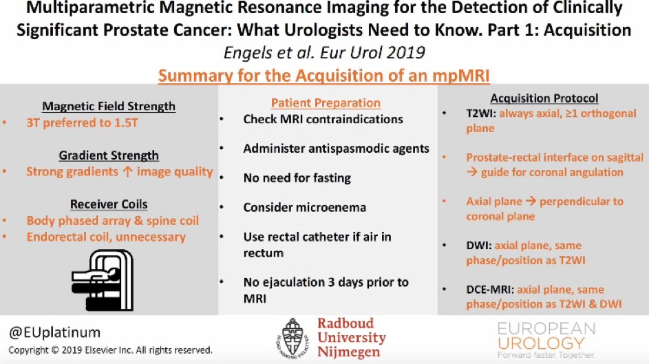
The success of multiparametric magnetic resonance imaging (mpMRI) for reliably detecting and localizing clinically significant (cs) prostate cancer (PCa) is highly dependent on image quality. However, due to the variability of available MRI equipment including software levels and prostate MRI technologists’ (MRI radiographers’) experience, it can be challenging to consistently achieve good-quality images for detection, localization, staging, and follow-up of PCa. The first step to improve quality and reduce variability is to implement optimized acquisition protocols.
Although the Prostate Imaging Reporting and Data System guidelines (2012 and 2016) describe minimal and optimal acquisition protocols for prostate mpMRI, these standards are not always met in daily practice. A major challenge in mpMRI is to obtain high image quality and reduce its variability for radiologic interpretations. A summary of evidence and guidelines for the acquisition of mpMRI of the prostate can set a basic guideline to reduce these variabilities (table 1).
Written by: Rianne R M Engels, Bas Israël, Anwar R Padhani, Jelle O Barentsz
Radboud University Nijmegen Medical Center, Nijmegen, The Netherlands., Paul Strickland Scanner Centre, Mount Vernon Hospital, Northwood, UK., Department of Radiology, Radboud University Nijmegen Medical Center, Nijmegen, The Netherlands.


